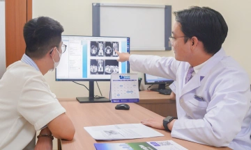
Anh Dat suffered from congenital atrophy of his left eyeball. His right eye, which was myopic, suddenly became blurry. Examinations at Tam Anh General Hospital Hanoi revealed his right eye’s vision had severely deteriorated, allowing him to only distinguish a hand held close to his face. Wide-angle fundus imaging confirmed retinal detachment extending to the macula—the central area responsible for detailed vision—accompanied by a tear. Based on his history of severe myopia and the characteristics of the damage, doctors diagnosed retinal detachment with a tear in his right eye due to severe myopia.
According to Doctor Bui Viet Hung, Head of the Vitreoretinal Department, severe myopia not only causes blurred vision but also significantly increases the risk of retinal detachment. As the eyeball lengthens over time, the retina stretches and thins, especially in the periphery, making it prone to degeneration and tears. Vitreous fluid can then penetrate these tears, causing the retina to separate from the underlying nourishing tissue and leading to detachment. This mechanism explains Anh Dat’s complication.
Since the retina had detached past the macula, there was an elevated risk of irreversible damage to photoreceptor cells if treatment was delayed, which would reduce the potential for vision recovery after surgery. "Without timely intervention, patients risk losing their sight completely," Doctor Hung stated.
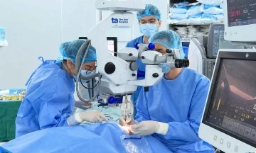
The surgical team decided to perform retinal detachment surgery to address the retinal tear, reattach it to its correct anatomical position, and facilitate maximum recovery of photoreceptor cells and preserve vision.
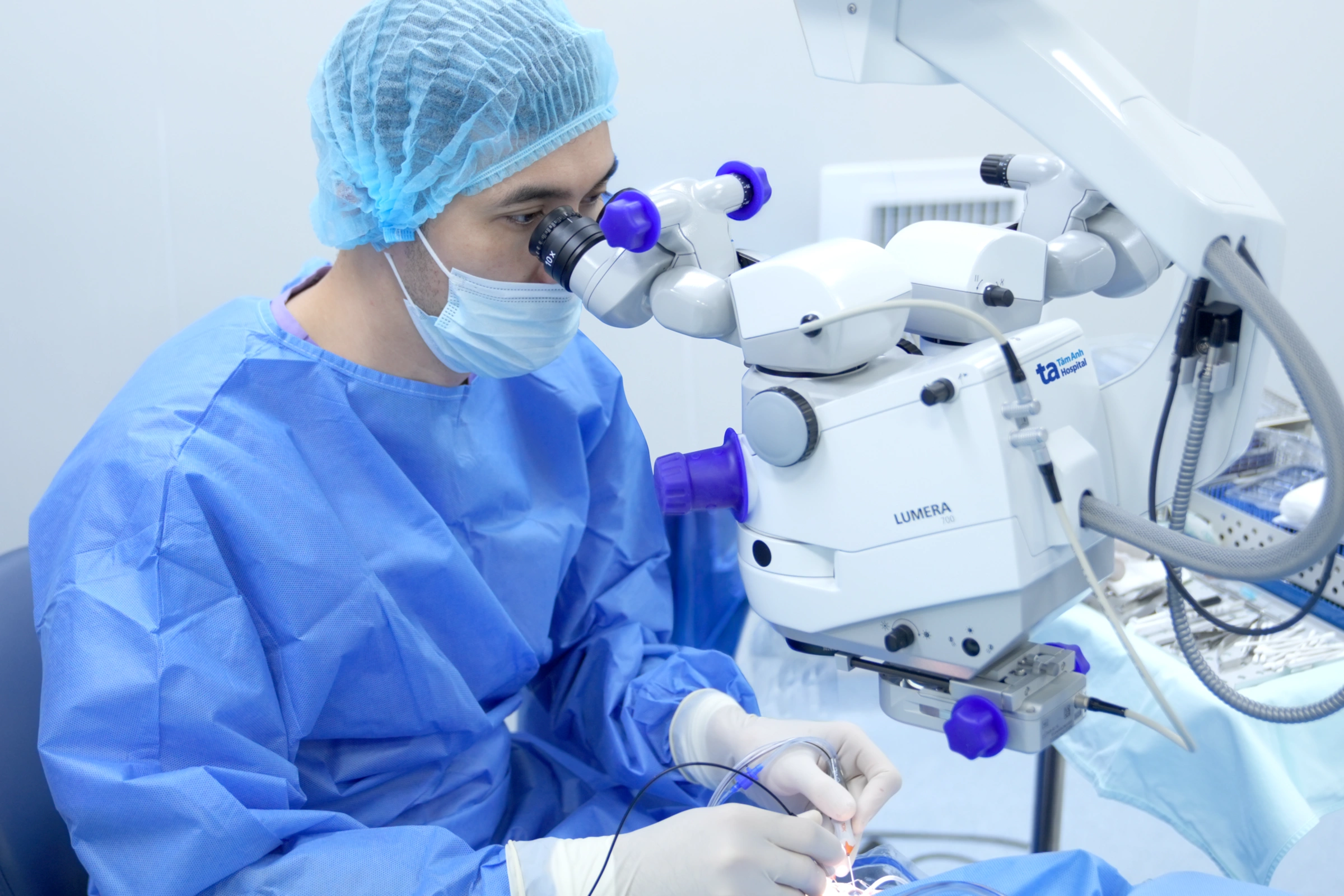 |
The surgical team performs retinal detachment surgery to preserve the patient's vision. *Photo: Tam Anh General Hospital*. |
Following surgery, Anh Dat’s vision improved significantly. He can now see, move around, and carry out daily activities independently.
Doctor Hung emphasized that severe myopic retinal detachment is an ophthalmological emergency where the timing of intervention is critical to treatment outcomes. When the retina is reattached promptly, photoreceptor cells have a better chance of recovery. Conversely, prolonged detachment due to delay leads to irreversible damage to these cells, drastically reducing the potential for vision improvement post-surgery.
Doctors advise myopic individuals, especially those with approximately 6 diopters or more or a long axial length, to undergo regular fundus examinations. This helps detect thin, degenerate, or torn areas of the retina early. If symptoms such as sudden blurry vision, flashes of light, numerous floaters, a sensation of a dark curtain obstructing vision, or distorted images appear, patients should seek immediate examination at a specialized eye clinic.
Thu Giang
| Readers can send questions about ophthalmic diseases here for doctors to answer. |