On 13/8, Doctor of Ophthalmology II Tran Ba Kien at VISI Eye Hospital reported that the patient's left retina was detached due to a tear, accompanied by degeneration in the peripheral retinal area. Her right eye also showed a tear and a degenerated retinal area, but no detachment yet.
"Retinal detachment caused by a long-standing tear can lead to vision loss, even irreversible blindness," the doctor stated.
The doctor applied two treatment methods for the patient's eyes. The left eye received intraocular gas injection, where a small gas bubble was introduced into the eye to press the retina back into its original position, combined with laser photocoagulation around the tear. For the right eye, the doctor used a laser to treat the weakened retinal area, aiming to reduce the risk of further damage leading to retinal detachment.
After three days, the patient's left retina had returned to its position and stabilized. With continued laser treatment, her vision gradually improved. The patient was advised to undergo regular follow-ups to assess recovery and detect any abnormalities early.
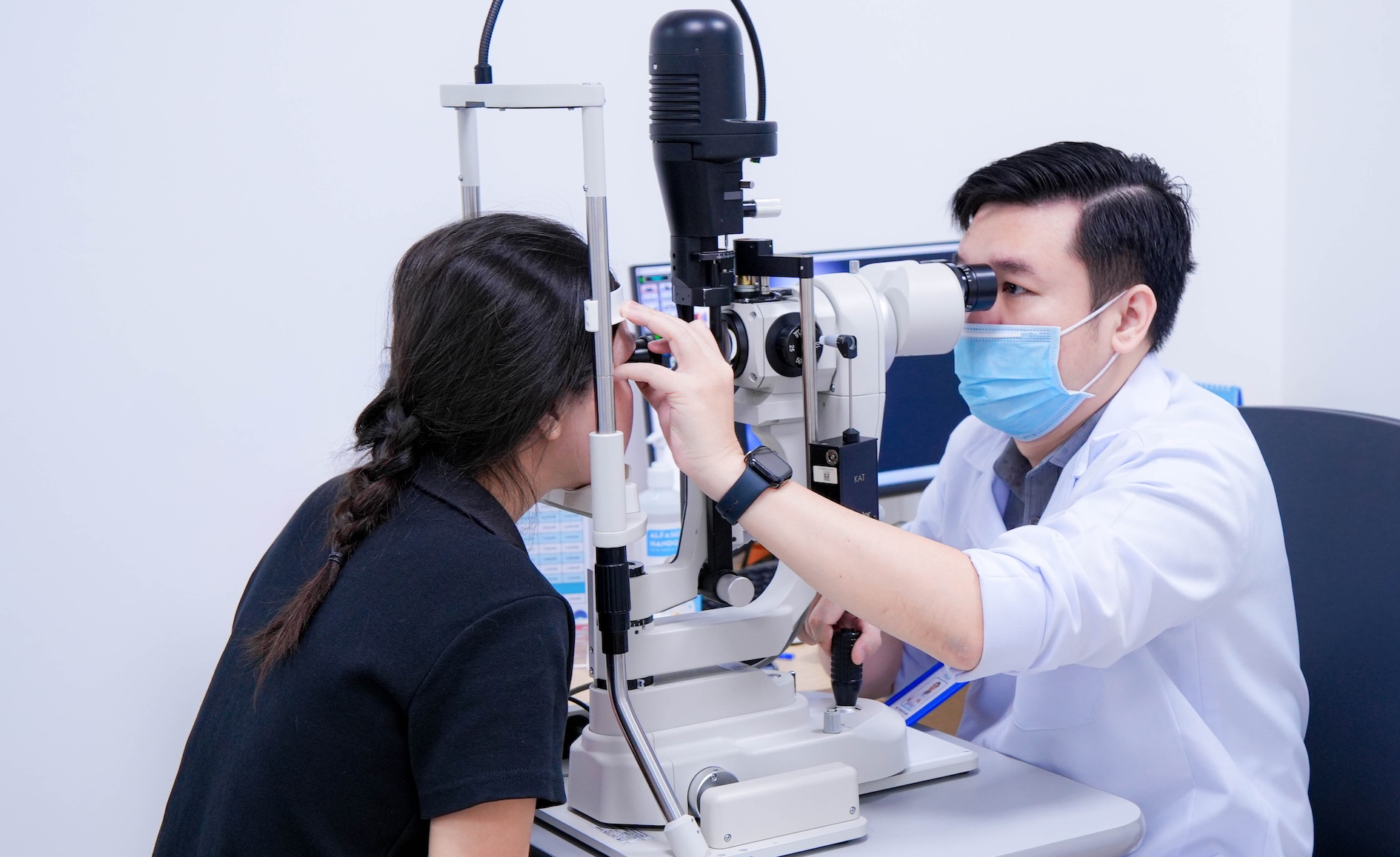 |
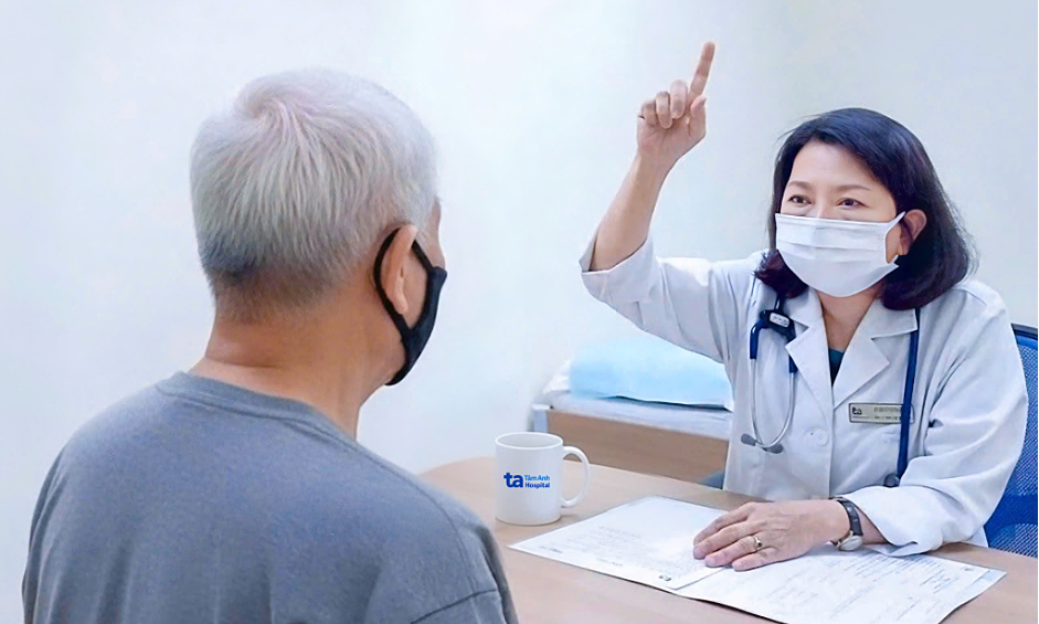
A doctor examines a patient's eye. Photo: Hospital provided.
Retinal detachment can be treated by various methods, including intraocular gas injection, vitrectomy, or scleral buckling. The choice of method depends on the location, number, and characteristics of the tear, the extent of the detachment, the condition of the vitreous, and individual patient factors.
According to Doctor Kien, myopic individuals, especially those with moderate and severe myopia, face a higher risk of retinal problems such as degeneration, tears, or detachment. In myopic people, the eyeball is typically longer, which can cause the retina to stretch and weaken. Severe myopia, particularly when the axial length of the eyeball exceeds 26 mm, is a risk factor for retinal detachment.
However, the degree of myopia alone cannot determine the risk for each person. For the patient mentioned, her myopia was only about 3 diopters. Risk also depends on the retinal condition, a history of holes or tears, previous retinal detachment in the other eye, family history, eye trauma, or surgery.
The doctor recommends that myopic individuals, especially those with severe myopia, undergo regular fundus examinations to detect weak or abnormal retinal areas early. If there is a sudden increase in 'floaters', continuous flashes of light, or a sensation like a dark curtain obscuring part of the vision, an eye examination is necessary immediately. These could be warning signs of a retinal tear or detachment.
Le Phuong