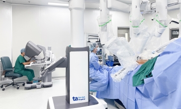
Dr. Pham Thanh Truc, a specialist from the Urology - Nephrology - Andrology Center at Tam Anh General Hospital, Ho Chi Minh City, reported that Mr. Ngoc was admitted with severe edema, fatigue, and altered consciousness. Tests revealed his glomerular filtration rate (eGFR) had dropped below 15 mL/min/1.73m², and ultrasound showed grade 3 hydronephrosis in both kidneys. Doctors diagnosed him with acute kidney failure.
A CT scan revealed bilateral ureteral stones. The left ureteral stone, measuring 3 cm, caused complete obstruction, deforming the ureteral tract into a Z-shape. Additional ureteral stones and a stone at the renal pelvis junction were also present. This blockage entirely prevented urine flow from the kidneys to the bladder, leading to acute kidney failure and infection.
Dr. Truc explained that immediate lithotripsy risked massive bacterial and toxin entry into the bloodstream through mucosal injuries, potentially causing septic shock, multi-organ failure, and life-threatening complications. The medical team opted to insert a JJ stent to drain urine, deferring immediate stone removal. This stabilized the acute kidney failure and infection, reducing pressure and ensuring safety for lithotripsy later.
One week after JJ stent insertion, Mr. Ngoc's blood and urine tests stabilized. The medical team used a flexible endoscope to perform bilateral kidney and ureteral lithotripsy, successfully clearing all stones in a single procedure.
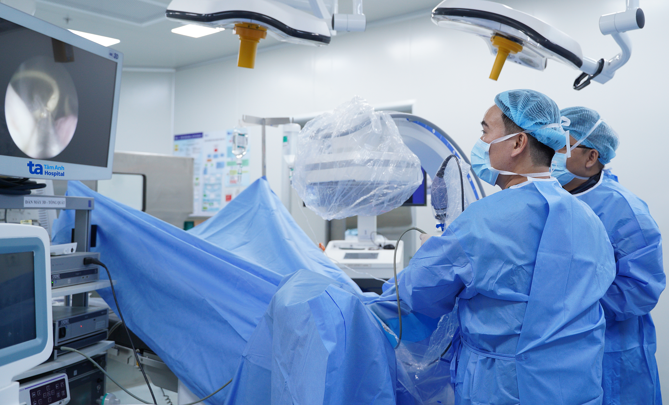 |
Dr. Truc (left) and the team perform percutaneous kidney stone lithotripsy for the patient. *Photo: Tam Anh General Hospital*
To optimize surgical outcomes, the team utilized a C-arm system for precise stone localization. This technology also allowed for immediate checks for residual stones post-procedure. Compared to open surgery, this minimally invasive method preserves healthy tissue, reduces blood loss, and lessens post-operative pain.
Following surgery, Mr. Ngoc's health stabilized, and kidney function recovered, with his glomerular filtration rate returning to a normal 85 mL/min/1.73m². His edema and fatigue resolved, leading to discharge after one day. Doctors advised regular follow-up for the removal of the two JJ stents.
Stone analysis revealed carbonate apatite and calcium oxalate monohydrate. Doctors advised the patient to increase water intake, limit high-protein and oxalate-rich foods: such as beets, chocolate, and various nuts. They also recommended medication to control urine pH and anti-infection agents. These measures, combined with lifestyle adjustments, aim to prevent recurrence. Given his susceptibility, Mr. Ngoc requires regular check-ups for early detection and intervention should new stones form.
Dr. Truc urges regular general health check-ups and abdominal ultrasounds for early detection of urinary tract stones, even before symptoms emerge. Patients should avoid complacency or self-treating with folk remedies, which can worsen the condition. Improper intervention risks stone displacement, mucosal damage, bleeding, or widespread infection, complicating and increasing treatment costs.
Bao Anh
*Patient's name has been changed