At 34 weeks of pregnancy, Nguyen Anh Tuyet, 27, from Hanoi, attended Hong Ngoc General Hospital for a routine check-up. During a specialized Doppler ultrasound, doctors discovered a fetal hepatic vascular malformation, specifically a congenital portosystemic shunt type III, along with polyhydramnios.
Doctor Do Danh Truong, a specialist in obstetrics and gynecology, explained that Tuyet's condition is a rare abnormality. While not always clinically evident, it can increase cardiac strain and affect fetal development.
"Without close monitoring, this hepatic vascular malformation could lead to fetal heart failure, fetal hydrops, or hemodynamic disturbances," the doctor stated.
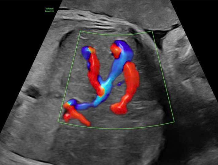 |
Ultrasound image showing the abnormal hepatic vascular malformation. *Hong Ngoc General Hospital* |
Following the detection of the anomaly, the team at Hong Ngoc General Hospital initiated a high-risk monitoring protocol for the pregnant woman, involving collaboration between obstetricians and imaging specialists. Tuyet's examination schedule became more frequent than a typical pregnancy, with check-ups occurring approximately once a week to continuously assess fetal status.
Each examination included a comprehensive fetal assessment via specialized Doppler ultrasound to evaluate blood flow within the hepatic vascular system, measuring the velocity and direction of blood through the abnormal shunt. Doctors also closely monitored hemodynamic indicators, such as cardiac output, signs of circulatory overload, and the risk of fetal heart failure.
"Beyond detecting the abnormality, we closely monitored its progression," explained Doctor Truong. "The malformation could remain stable, worsen, or, rarely, improve on its own. Early detection of warning signs, such as an enlarged fetal heart, abnormally increased blood flow, or fluid retention, is crucial for timely intervention."
In parallel with hospital monitoring, doctors maintained contact with Tuyet, instructing her on how to recognize signs like fetal movement and abdominal distension due to polyhydramnios, enabling her to seek prompt care if abnormalities arose. Five weeks later, the fetal condition remained stable, with no signs of heart failure or severe complications. However, given the potential for rapid progression of vascular malformations, the team continuously prepared appropriate intervention plans.
At 39 weeks of pregnancy, after examination and rapid assessment of indicators, doctors observed signs that the fetus required early intervention to prevent complications during labor. Following a consultation, the team decided on a Cesarean section.
"For cases with circulatory abnormalities like hepatic vascular malformations, choosing the birth timing is critical," Doctor Truong elaborated. "Prolonging the pregnancy too much can increase risks for the fetus, while intervening too early, before the fetus is mature enough, is also not beneficial. We must carefully consider all factors to make the optimal decision."
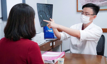
 |
Routine prenatal check-ups help pregnant women monitor the health of both mother and baby, allowing for timely management of abnormalities. *Hong Ngoc General Hospital* |
The surgery proceeded smoothly, and the baby was born stable, with no signs of respiratory distress or circulatory disorders. Immediately afterward, the infant was transferred to the Neonatal Unit for continued close monitoring and was discharged after three days with a schedule for follow-up appointments. Doctor Truong reported that after five months, the baby's hepatic vascular malformations had shrunk and gradually closed, with all developmental indicators and liver functions within normal ranges, demonstrating healthy growth.
The doctor emphasized that fetal hepatic vascular malformation is a rare condition, typically detected only through specialized Doppler ultrasound using high-tech equipment and experienced medical professionals. Depending on its severity, some cases may not cause serious effects, but if the lesion is large, the child risks complications such as heart failure, fetal hydrops, anemia, or coagulation disorders.
"Routine prenatal check-ups play a pivotal role in the early detection and effective management of abnormalities," added Doctor Do Danh Truong. "With systematic monitoring, doctors can proactively manage risks, choose appropriate intervention times, and plan postnatal follow-up, ensuring the safety of both mother and baby."
Hieu Chau
Department of Obstetrics and Gynecology - Hong Ngoc General Hospital Phuc Truong Minh
Address: 2nd floor, 8 Chau Van Liem, Tu Liem, Hanoi
Hotline: 0919 645 271 - 0918 750 845