Ms. Nhu, a 28-year-old woman, successfully underwent robot-assisted surgery to remove a 3 cm pancreatic mucinous cyst at Tam Anh General Hospital, TP HCM. The procedure prevented the lesion from potentially developing into cancer, offering her a positive prognosis.
Initially, an ultrasound at the hospital revealed a cyst-like structure measuring approximately 33 mm on her pancreatic body, adjacent to the left side of the abdominal aorta. Subsequent MRI scans clearly showed the cyst located behind the main pancreatic duct but not connected to it, with no dilation of the duct or fluid around the pancreas.
Doctor Pham Cong Khanh, Head of the Department of Hepatobiliary-Pancreatic Surgery at the Center for Endoscopy and Endoscopic Digestive Surgery, diagnosed the lesion as a mucinous cystic neoplasm (MCN). MCN is a precancerous pancreatic cyst that can progress to malignancy. Early detection and removal of such lesions are crucial for preventing cancer development.
The lesion's location, behind the pancreatic duct and near vital blood vessels like the splenic artery and vein, presented a complex surgical challenge. Following a consultation, the medical team opted for surgery using the Da Vinci Xi robot system.
The robot provided high-resolution, three-dimensional images of the surgical field, enabling doctors to clearly visualize structures deep within the abdominal cavity. Its flexible arms allowed for precise dissection, minimizing damage to critical blood vessels.
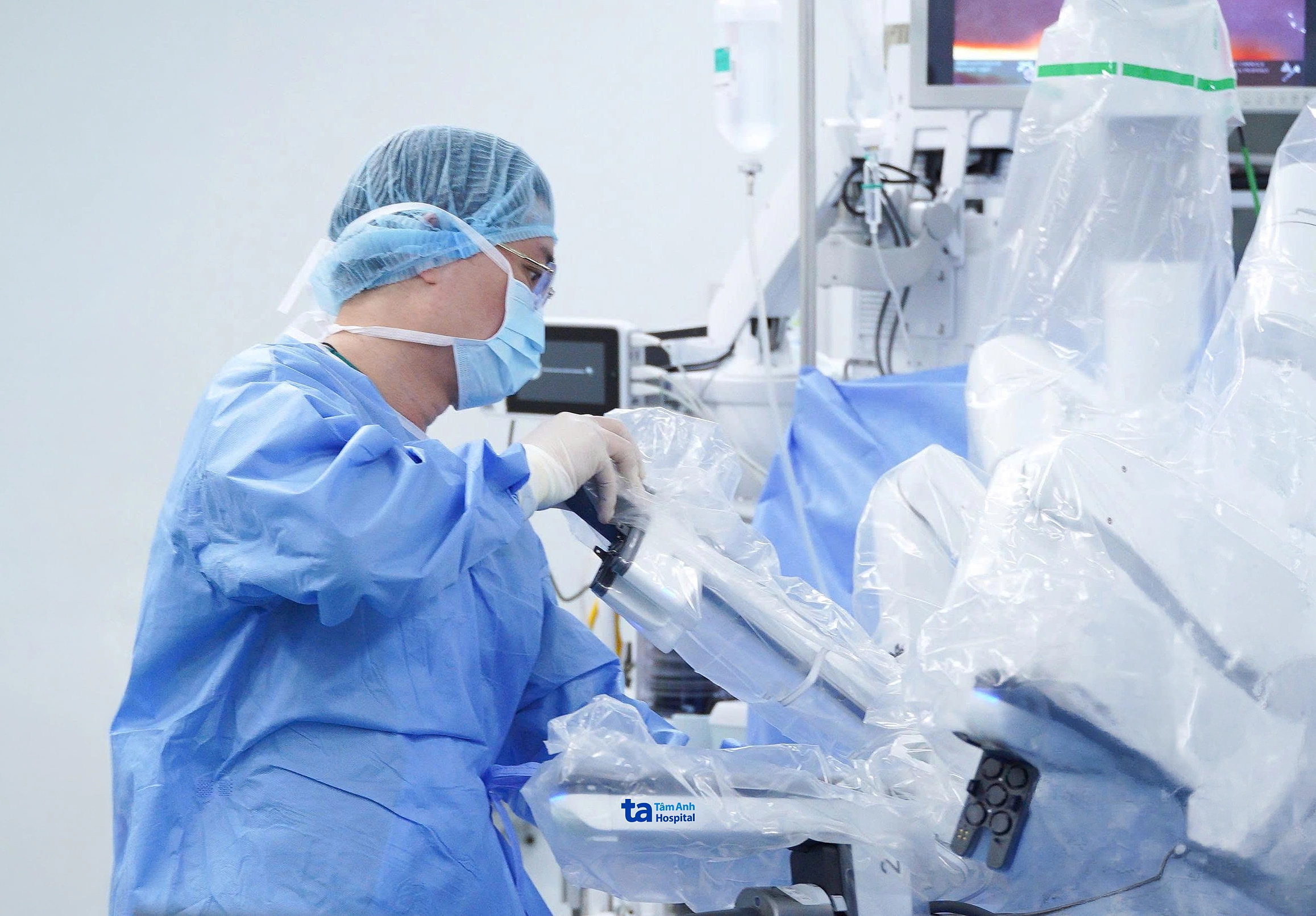 |
Doctor Khanh attaches the robot arm to remove Ms. Nhu's pancreatic cyst. Photo: Tam Anh General Hospital |
During the operation, the team accessed the omental bursa region and found the cyst, measuring about 30 mm, located on the upper border of the pancreas, behind the splenic artery, with increased vascularity around it. Surgeons carefully dissected the lower border of the pancreas from the body to the tail, then identified the splenic artery on the upper border. The pancreatic body was transected about 1 cm from the lesion's edge. The entire body and tail of the pancreas, along with the cyst, were mobilized, while preserving the splenic vascular bundle and the spleen.
Post-surgery, Ms. Nhu experienced no complications such as pancreatic leakage or bleeding and was discharged after 5 days. Pathological results confirmed the lesion as a mucinous cystic neoplasm, with clear margins, indicating no abnormal cells remained.
 |
Ms. Nhu receives care from a nurse after surgery. Photo: Tam Anh General Hospital |
Doctor Khanh emphasized that pancreatic cysts often present without symptoms, leading to their incidental discovery during routine health check-ups. Abdominal ultrasound is often the initial screening method. However, due to the pancreas's deep location within the abdomen and potential image limitations from intestinal gas, further evaluation with CT, MRI/MRCP, or endoscopic ultrasound may be necessary for some lesions.
Doctors advise individuals to seek medical attention for persistent symptoms such as: pain in the epigastric region or left hypochondrium, radiating to the back, persistent bloating, indigestion, loss of appetite, or unexplained weight loss. Early detection of pancreatic cysts allows doctors to monitor, assess risk, and intervene appropriately, preventing lesions from progressing to cancer.
Quyen Phan
| Readers can submit questions about digestive diseases here for doctors to answer |