Ultrasound and gastroscopy at Tam Anh General Hospital, Ho Chi Minh City, revealed a 15 mm tumor in Ms. Lan's left kidney. She had no history of high blood pressure, diabetes, or urinary dysfunction.
Dr. Nguyen Tan Cuong, Deputy Head of the Urology Department and Head of the Urological Cancer Unit at the Urology - Nephrology - Andrology Center, stated that the tumor was located in one-third of the kidney, near the renal hilum and vascular system. It was suspected to be early-stage malignant due to strong contrast enhancement. The optimal treatment was laparoscopic partial nephrectomy to remove cancer cells and preserve kidney function.
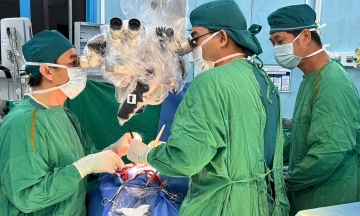
However, Ms. Lan's tumor was close to major blood vessels, requiring precise dissection to prevent massive bleeding or permanent kidney damage. Therefore, doctors opted for Da Vinci Xi robotic surgery.
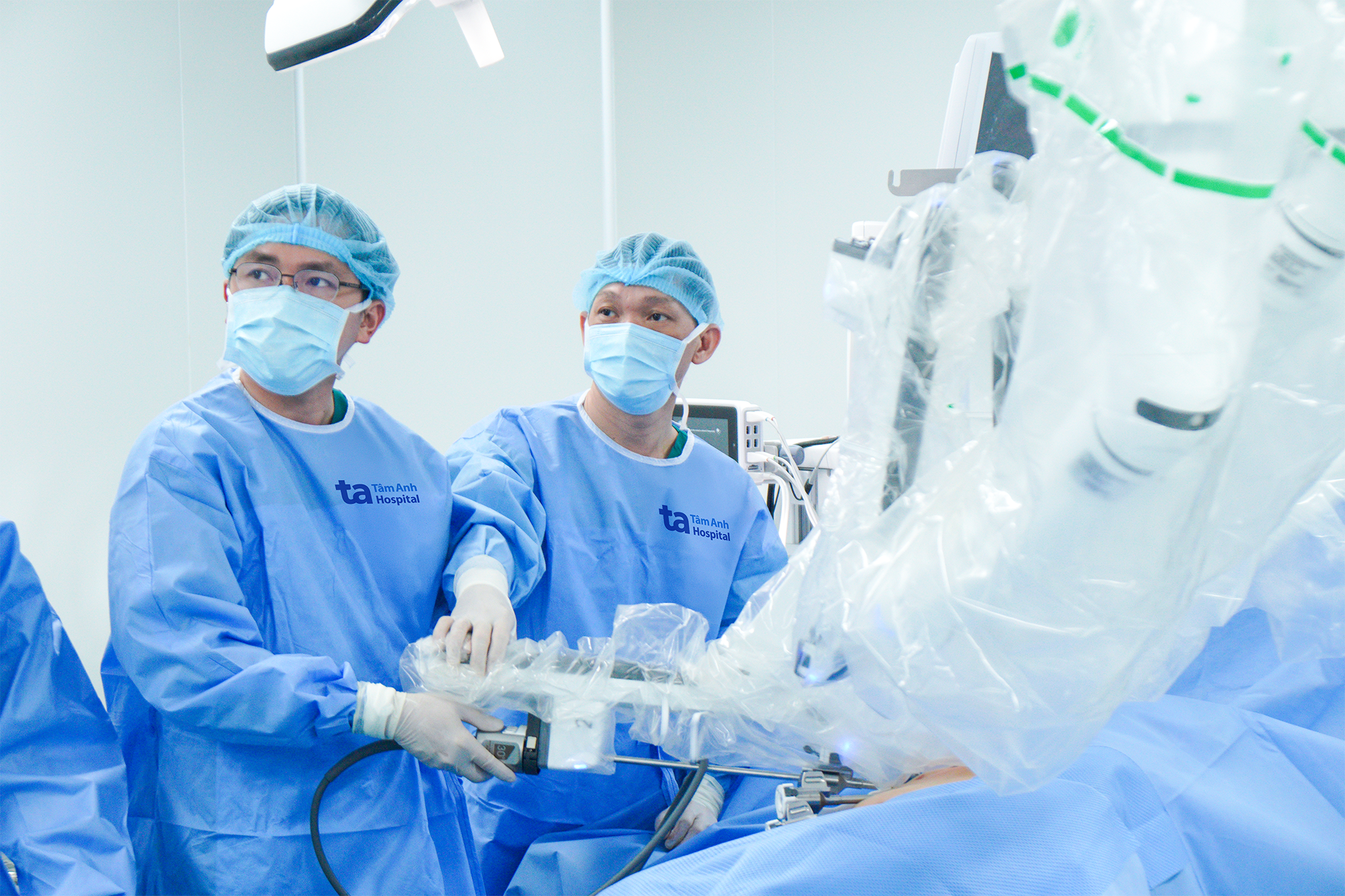 |
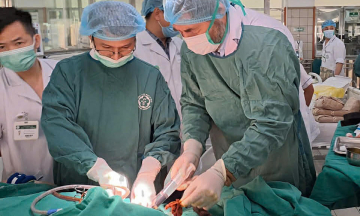
Dr. Cuong (right) prepares before controlling the robotic arms to remove Ms. Lan's kidney tumor. Photo: Tam Anh General Hospital |
Using the magnified 3D camera system and flexible robotic arms, Dr. Cuong clearly observed capillaries and tumor boundaries, allowing for precise dissection and rapid kidney tissue suturing.
The team performed the tumor removal without clamping the renal artery, preventing post-operative kidney function impairment. Robotic surgery allowed doctors to remove the tumor, control bleeding, and preserve kidney function.
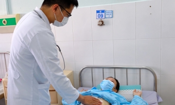
Ms. Lan recovered quickly. One day after surgery, she could sit up, eat, and walk gently, experiencing minimal incision pain. After 3 days, her vital signs were stable, and she was discharged without needing additional chemotherapy or radiation.
Dr. Cuong noted that kidney tumors often result from genetics, environmental factors, chemicals, or lifestyle. Early-stage kidney cancer typically lacks clear symptoms, emphasizing the need for healthy living and regular health screenings. Early detection via ultrasound or CT scans improves treatment success rates and preserves kidney function.
Bao Anh
* Patient's name has been changed