The baby girl was diagnosed with a sacrococcygeal tumor, located at the end of the spine just above the gluteal cleft, during a fifth-month prenatal ultrasound. By the 37th week of pregnancy, doctors noted polyhydramnios and rapid tumor growth with grade two vascular proliferation, indicating a risk of premature rupture of membranes and preterm birth. The mother had no underlying medical conditions, a normal pregnancy, and no family history of this condition. Doctors advised regular prenatal check-ups for close monitoring of the fetus.
The baby was born via natural delivery at 39 weeks, weighing 3,7 kg, breathing independently, and feeding well. However, the large sacrococcygeal tumor presented risks, leading the family to transfer the baby to Tam Anh General Hospital Ho Chi Minh City. Dr. Le Thi Ngoc Dung, from the Neonatal Center, diagnosed the baby with a type two sacrococcygeal teratoma. The tumor was primarily external, with a small portion attached to the posterior-inferior aspect of the sacral vertebrae, measuring approximately 86x80x97 mm.
Before surgery, the baby received special care to ensure adequate energy. Doctors closely monitored her heart rate, breathing, blood pressure, circulatory status, and managed the risk of bleeding from the tumor. Concurrently, tests were conducted to evaluate heart and lung function, blood coagulation, and imaging to determine the extent of tumor invasion, facilitating the selection of a safe anesthesia and surgical plan.
Dr. Nguyen Do Trong, from the Pediatric Surgery Department, explained that the large tumor had a complex location and a mixed composition of solid and cystic components with multiple septa, forming a honeycomb structure. This indicated a high risk of malignancy. The tumor occupied a significant portion of the subcutaneous tissue of both buttocks, making contact with the gluteus maximus muscle, the posterior rectal wall, and the coccyx. Dr. Trong decided on early surgical removal to prevent complications such as tumor rupture, infection, and tissue necrosis.
The airway of a three-day-old neonate is very small, and the respiratory and circulatory systems are immature, making them vulnerable to functional impairment. Consequently, doctors precisely calculated the anesthesia procedures and medication dosages. The medical team closely monitored vital signs, ready to intervene immediately if hypoglycemia, hypothermia, electrolyte imbalance, or sudden apnea occurred. A ventilator, warming machine, emergency medications, and intravenous fluids were all prepared.
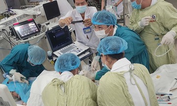
After three hours, the team completely removed the tumor, which was then sent for histopathological analysis. They also performed perineal reconstruction, preserving adjacent anatomical structures. The baby received 70 ml of blood to compensate for blood loss, maintaining stable vital signs.
Post-surgery, the baby's vital signs continued to be closely monitored, and she received medication and breast milk feeding in the Neonatal Intensive Care Unit (NICU). After two days, the incision was dry, and the baby was feeding well, leading to discharge. Histopathology results indicated a suspected malignant teratoma, necessitating ongoing monitoring by pediatric oncology specialists.
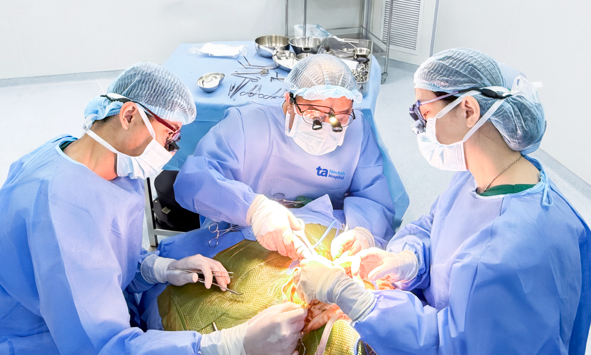 |
Dr. Trong (center) and the surgical team removing the tumor from the baby at three days old. Photo: Tam Anh General Hospital |
Dr. Trong stated that sacrococcygeal teratomas typically develop from primitive germ cells and appear in the coccygeal region. The exact cause of these teratomas remains unclear.
Sacrococcygeal teratomas are classified into four types based on their location and extent of invasion. Type one tumors are entirely external. Type two, like this baby's case, are primarily external but have a small portion attached to the coccyx or vertebrae. Type three tumors are mostly located within the abdomen and pelvis, with only a small external protrusion that may contact various internal organs. Type four tumors are entirely contained within the abdomen or true pelvis. Dr. Trong cited research indicating that the incidence of sacrococcygeal teratomas in neonates is approximately 1/35.000 to 1/40.000 live births, occurring more frequently in baby girls. Of these, about 7-10% of cases are malignant teratomas.
Doctors advise pregnant women to undergo all scheduled tests and regular ultrasounds during prenatal check-ups. Early detection of abnormalities during pregnancy helps doctors plan for timely monitoring and intervention.
Gian Don
| Readers can submit questions about neonates here for doctors to answer. |