PET/CT combines two imaging techniques: positron emission tomography (PET), which provides early molecular-level images, and computed tomography (CT), which offers clear anatomical views. This combination aims to detect and assess the malignancy and spread of tumors, lymph nodes, and distant metastatic lesions earlier than conventional imaging methods.
Doctor Vu Huu Khiem, head of the Oncology Department at Tam Anh General Hospital Hanoi, explained that during a PET/CT scan, patients receive an intravenous injection of a mildly radioactive substance, typically fluorodeoxyglucose (FDG). FDG is a glucose-like molecule that all body cells use for fuel, but it is tagged with Fluor-18 to act as a tracer.
In the body, cells absorb FDG just as they would glucose. Cancer cells require more energy for proliferation and growth, leading them to absorb higher amounts of glucose and FDG. PET/CT utilizes sensors to pinpoint locations with increased positron emissions, which correspond to the sites of primary and metastatic cancer cells.
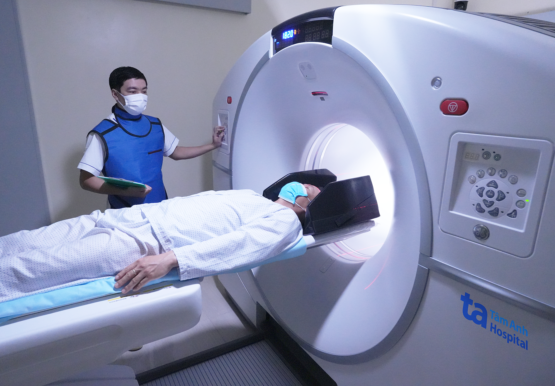 |
A patient undergoing a PET/CT scan. Photo: Tam Anh General Hospital.
Doctors prescribe PET/CT for cancer patients in several situations:
To distinguish between benign and malignant tissue: When other imaging techniques, such as computed tomography (CT) and magnetic resonance imaging (MRI), only reveal the morphology of a lesion, like a mass or nodule, without clearly indicating whether it is benign or malignant, a PET/CT scan enhances the ability to differentiate more accurately by evaluating the tumor's characteristics. This can reduce the need for unnecessary biopsies.
To locate the primary cancer site: When doctors suspect cancer based on blood tests and clinical symptoms but are unsure of the tumor's location, a whole-body PET/CT scan helps identify the suspected primary cancer site, supporting biopsy for disease confirmation.
To stage cancer: PET/CT also assists doctors in determining tumor size and whether the cancer has metastasized to lymph nodes or other organs. The whole-body scanning capability of PET/CT allows for the detection of small metastatic lesions that other methods might overlook.
For treatment planning: This method helps doctors accurately identify the location and extent of the developing cancer. This information is crucial for planning surgery or radiation therapy, ensuring that radiation beams are precisely aimed at the tumor, reducing the risk of missing lesions, preventing recurrence, and limiting damage to surrounding healthy tissues.
To evaluate treatment effectiveness: During chemotherapy, radiation therapy, targeted therapy, or immunotherapy, doctors may order a PET/CT scan to assess the tumor's response to treatment.
To monitor for cancer recurrence: After treatment is completed and the patient is in complete remission or cured, doctors may prescribe PET/CT during routine follow-up appointments to check for cancer recurrence, allowing for timely intervention. PET/CT detects lesions at the molecular level, often several months earlier than other conventional techniques.
PET/CT scans are not indicated for pregnant women due to the potential impact of CT radiation on the fetus. Breastfeeding women who require a PET/CT scan should suspend breastfeeding for 24 hours post-scan. Patients with a history of contrast dye allergy or kidney failure can undergo PET/CT without contrast.
Doctor Khiem notes that PET/CT scans are not recommended for cancer screening due to the risk of false positives and high costs. Images showing strong FDG metabolic activity can also indicate infection or inflammation, not necessarily cancer. A definitive cancer diagnosis ultimately relies on biopsy results for histopathological examination. Patients should only undergo PET/CT when specifically indicated by a specialist, after a careful consideration of the clinical benefits outweighing the risks and costs.
By Thanh Long
| Readers can ask questions about cancer here for doctors to answer. |