Retinopathy of prematurity (ROP) is a condition where retinal blood vessels develop abnormally in premature infants whose retinal vascular system is not yet complete. ROP can range from mild, self-resolving cases to severe damage that significantly impacts vision, making timely screening crucial.
Not all premature babies require ROP screening, and there is no single guideline for every infant. According to the 2024 updated guidelines from the Royal College of Paediatrics and Child Health, the primary criteria are infants born at less than 31 weeks gestational age or with a birth weight under 1,5 kg. The guidelines also recommend considering screening for infants older than 31 weeks or those with unstable health and a high risk of ROP. The timing of the first eye exam depends on gestational age at birth, postnatal age, and postnatal progression, such as respiratory status and infections.
Doctors assess retinal blood vessel development through a specialized fundus examination. Some facilities may use wide-field digital retinal imaging systems to record images and monitor changes across examinations. Exam results describe the retinal zone, ROP stage, and signs of abnormal blood vessels. This information helps doctors determine when follow-up examinations are needed.
The interval between examinations is not fixed. Many infants have follow-up exams after a few weeks; however, babies with immature retinas or signs of ROP progressing to severe stages requiring laser intervention or intravitreal injections are monitored more closely.
ROP eye examinations can cause discomfort and pain; infants may cry or show changes in physiological parameters during the exam. Therefore, comfort-reducing measures such as topical anesthetics, sucrose solution, pacifiers, or swaddling may be used depending on the case.
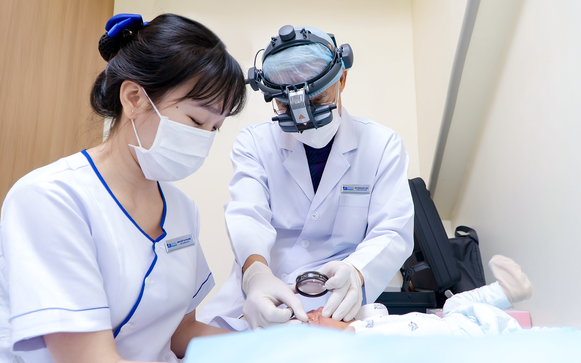 |
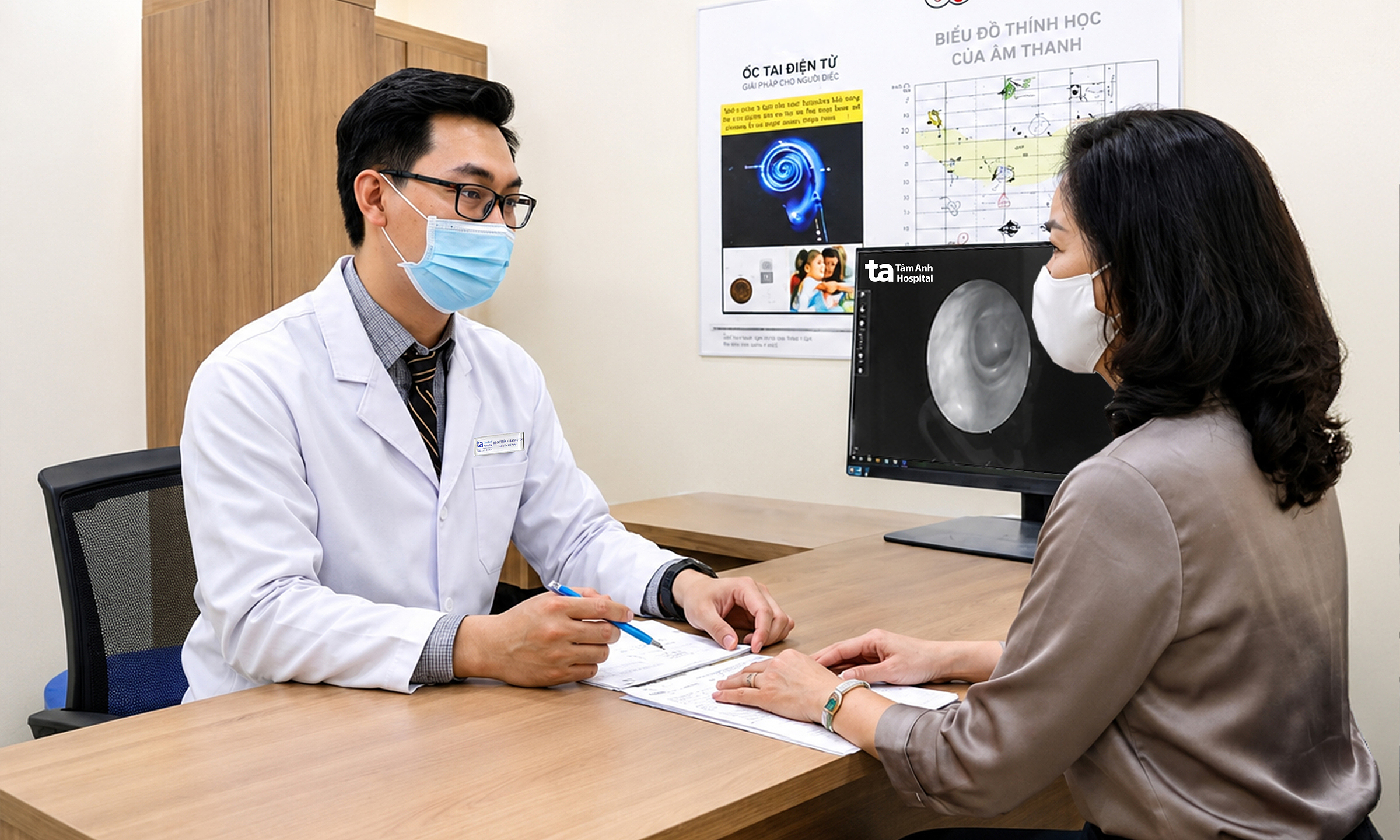
Premature babies undergo retinopathy of prematurity (ROP) screening to prevent the risk of vision damage. Illustration: Tam Anh General Hospital |
Each premature baby typically undergoes 3-6 eye examinations on average, potentially more for extremely premature infants or those with severe complications. The examination schedule continues until the retinal blood vessels are fully developed or the ROP lesions safely regress. ROP can progress rapidly, making timely follow-up appointments essential. Premature babies face a higher risk of certain vision problems than full-term infants, so screening as directed by a doctor is crucial.
ROP screening can be performed collaboratively by neonatologists and ophthalmologists, allowing for monitoring of the infant's retinal condition while in the incubator in the Neonatal Intensive Care Unit (NICU). With wide-field retinal imaging systems, fundus images can be recorded and stored, enabling doctors to compare and assess progression across examinations.
The examination process requires strict infection control. In some cases, appropriate pain and discomfort reduction measures can be applied for the child during the examination.
Doctor Nguyen Minh Thanh Giang
Neonatal Center
Tam Anh General Hospital, TP HCM
| Readers can submit questions about pediatric diseases here to receive answers from doctors. |