Over one year, the patient's grip strength gradually declined, making seemingly simple actions like buttoning a shirt difficult. He reported no history of trauma, cervical spine pain, or hand numbness.
Doctors at Hospital 108 observed clear atrophy of the interosseous muscles and hypothenar eminence in the patient's left hand, along with forearm atrophy, predominantly on the ulnar side—all characteristic signs of Hirayama disease. Although the patient noticed no abnormalities in his right hand, doctors detected mild atrophy of the dorsal interosseous muscles. This subtle finding suggested that the damage might not be confined solely to the symptomatic hand.
Electromyography results indicated an anterior horn cell lesion, primarily affecting the C8-T1 spinal cord segments bilaterally, with the left side more severely affected than the right. The electromyogram also revealed abnormalities in the right hand, which the patient had not perceived as weak or significantly atrophied. Sensory responses remained preserved, consistent with the characteristics of Hirayama disease.
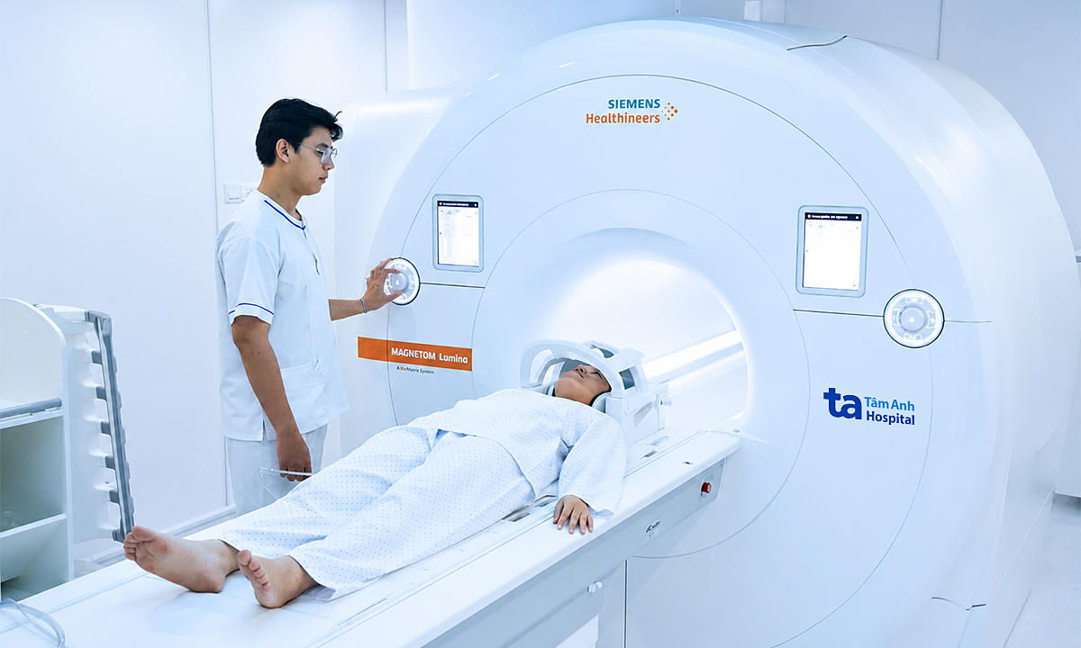
Magnetic resonance imaging of the cervical spine showed no abnormalities in the cervical spinal cord in a neutral position. However, an MRI performed in cervical flexion revealed anterior displacement of the posterior dura, widening of the epidural space, and compression of the lower cervical spinal cord. These are characteristic signs confirming Hirayama disease.
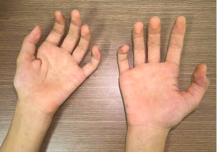 |
The left hand exhibits more pronounced muscle atrophy than the right hand. *Photo: Hospital provided*
Hirayama disease, also known as juvenile spinal muscular atrophy, was first described by Japanese scholar Keizo Hirayama and colleagues in 1959. It is a rare cervical myelopathy characterized by self-limiting, asymmetrical, and slowly progressive atrophy of the forearm and hand, primarily affecting young males aged 15 to 25.
There is currently no specific cure for Hirayama disease. If detected early, patients receive conservative treatment, which includes limiting excessive neck flexion, potentially using a cervical collar during the progressive phase, and incorporating physical therapy to maintain muscle strength and improve hand function. If the disease progresses rapidly, muscle weakness worsens, or conservative treatment proves ineffective, doctors may consider surgical cervical spinal cord decompression to alleviate pressure and limit dural displacement.
Dr. Nguyen Tuong Ngoc Linh, Senior Specialist and Deputy Head of the Department of Functional Diagnostics, stated that Hirayama disease typically progresses for about three to five years before entering a stable phase. Therefore, young individuals experiencing gradual, progressive weakness or atrophy in their hands or forearms, especially without sensory disturbances, should seek early medical attention. A combination of clinical examination, electromyography, and cervical spine MRI in a flexed position can detect the disease, enabling appropriate treatment, monitoring, and preservation of motor function for the patient.
Le Nga