Frederik was admitted to Tam Anh General Hospital Ho Chi Minh City for surgery following a fall two weeks prior. Doctor Le Dinh Khoa, head of the joint reconstruction department at the orthopedic trauma center, reported that the patient had a severely displaced fracture of the right femoral head. Hip muscle contracture had occurred due to limited movement, causing intense pain and an inability to walk independently. The surgery presented significant challenges due to the patient's tall stature and distinct anatomical joint structure compared to Asian individuals, necessitating the selection of an appropriately sized artificial joint. He also suffered from multiple underlying conditions, including hypertension and diabetes, which increased the risk of surgical complications.
Following a consultation, the medical team simultaneously employed several advanced 3D technologies to prepare for the operation. Doctors ordered a 3D CT scan, utilizing over 100,000 slices to generate a detailed three-dimensional image of the hip joint. This allowed for comprehensive observation of bone and joint structures and detection of potential lesions. This technology delivers a very low radiation dose, equivalent to a single X-ray, yet produces complete bone images, aiding in diagnosis and treatment.
Next, doctors simultaneously employed two specialized orthopedic software programs, TraumaCAD and MediCAD 3D, to enhance accuracy in selecting the artificial hip joint. TraumaCAD analyzes joint structure based on X-ray images, while MediCAD 3D formulates surgical plans using 3D imaging data from CT scans. These software solutions enable doctors to precisely locate anatomical structures, calculate acetabular inclination, determine artificial hip placement orientation, and simulate joint movements. This capability allowed doctors to select a joint suitable for the patient's body and develop a highly accurate surgical plan.
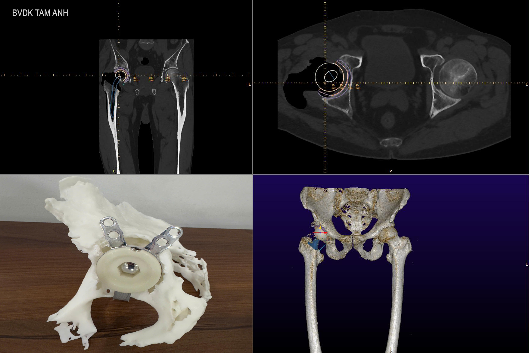 |
3D technologies used in Mr. Frederik's surgery: 3D CT scan, calculations on MediCAD 3D, and a 1:1 scale hip joint model. *Photo: Tam Anh General Hospital* |
Doctor Khoa conducted a "virtual" trial surgery, utilizing 3D printing technology to create a 1:1 scale hip joint model. This allowed the team to anticipate potential surgical scenarios, plan rapid responses, and ultimately shorten the actual operation time.
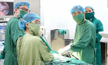
Doctors performed the hip replacement using the anterior based muscle sparing (ABMS) approach. This technique allows deep access to replace the hip joint without cutting muscles. Post-surgery, the muscles surrounding the joint remained intact, resulting in less pain, quicker recovery, and avoidance of risks such as dislocation, nerve damage, or post-operative paralysis. Patients can then perform complex movements after replacement, including squatting and crossing their legs.
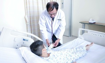
On the first post-operative day, Frederik experienced significant pain reduction, was able to stand and perform exercises with a walker, and had balanced leg lengths. At a follow-up appointment two weeks later, the patient had recovered almost completely and could walk independently without assistance.
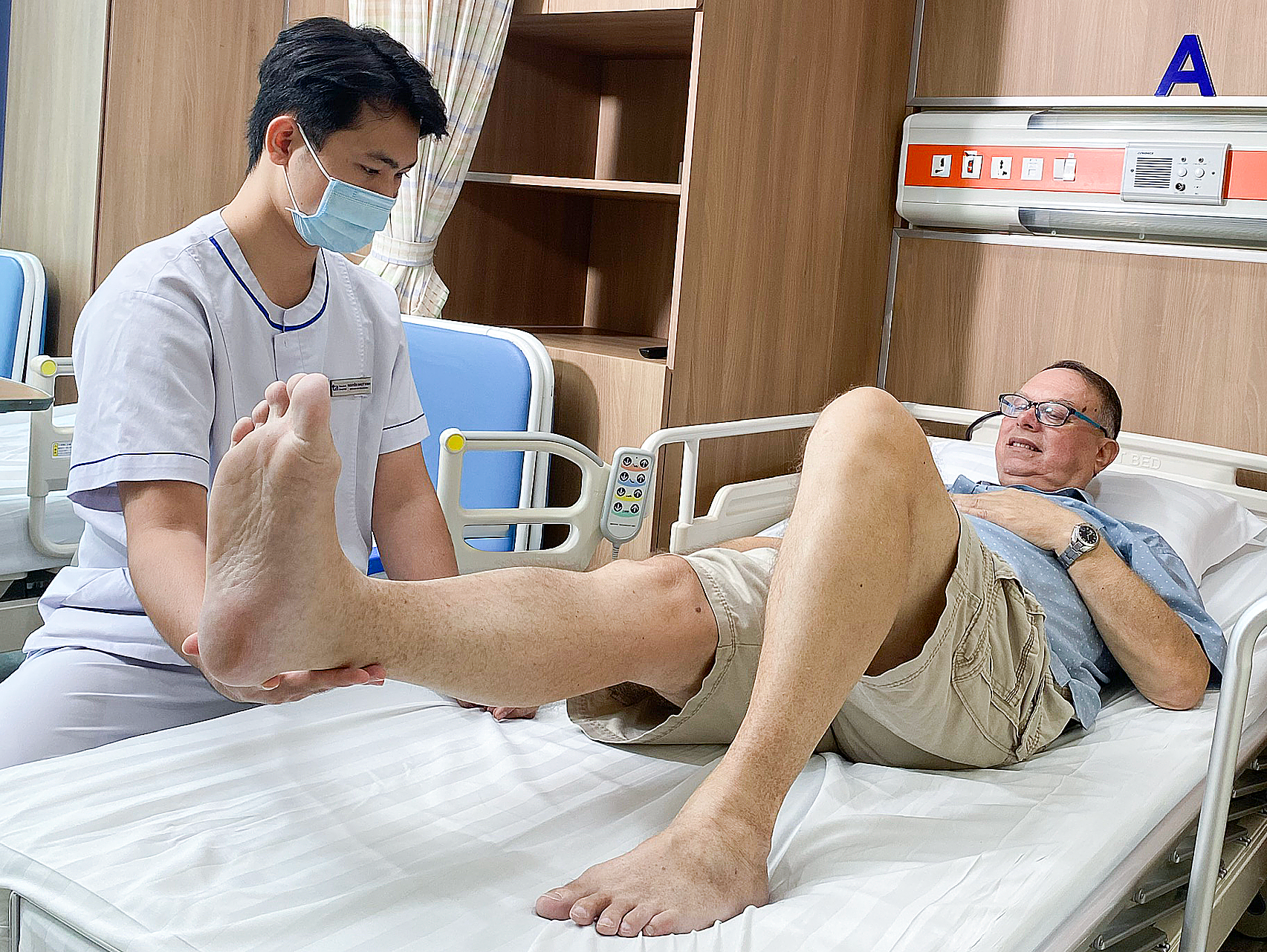 |
Frederik undergoing physical therapy on the second day post-surgery with a technician before discharge. *Photo: Tam Anh General Hospital* |
According to Doctor Khoa, the femoral neck is a unique structure located deep within the hip joint. It is susceptible to poor blood supply, making natural healing difficult after a fracture, particularly in older individuals. Surgery is often the optimal treatment method. Doctors advise that immediately after an injury, patients should seek examination at specialized musculoskeletal or orthopedic trauma hospitals for timely treatment and to prevent complications.
By Phi Hong