Dr. of First Degree Specialization Doan Thi Phuong Nhi, from the High-Tech Eye Center at Tam Anh General Clinic, District 7, reported that the girl's vision in both eyes was 10/10, with no pain. However, a slit-lamp microscope examination revealed conjunctival hemorrhage in the left eye and a poor pupillary light reflex. After administering pupil-dilating eye drops, a peripheral retinal examination identified a superior retinal tear, accompanied by vitreous hemorrhage and an extensive subretinal hematoma. Results from optical coherence tomography (OCT), color fundus photography, and ultrasound confirmed vitreous hemorrhage, iris sphincter rupture, and a peripheral retinal tear in the girl's left eye; her right eye was normal.
Dr. Nhi explained that the impact from the cable caused the eyeball to be compressed and then suddenly rebound, creating traction between the vitreous and the retina. This mechanism led to the retinal tear, particularly in the peripheral region, which is thin and vulnerable. Since the tear was located far from the center of vision, it might not have initially affected her eyesight. This often causes patients to be complacent, missing the optimal treatment window and only discovering the issue once retinal detachment has occurred.
"This is a severe injury; if not detected and treated promptly, the child faces a risk of retinal detachment, leading to permanent vision loss", Dr. Nhi stated.
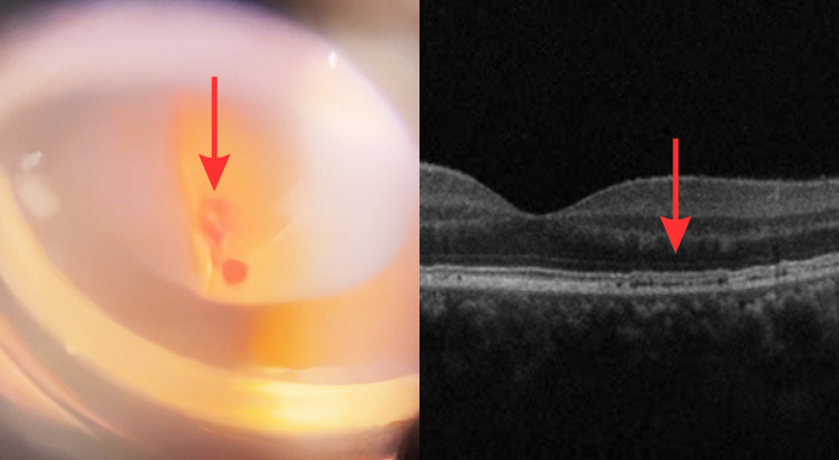 |
Hemorrhage in the left eye (left image) and OCT scan showing retinal pigment epithelial detachment in the left eye (right image). *Photo: Tam Anh General Hospital* |
The doctor prescribed eye drops and oral medication to reduce inflammation, support the absorption of blood from the vitreous hemorrhage, and treat the surface damage to the eyeball. Once the vitreous hemorrhage was stably controlled, the patient was scheduled for peripheral retinal laser photocoagulation around the tear. This procedure aims to seal the tear, prevent fluid leakage, and help preserve her vision.
One week after treatment, the retinal condition was stable, and the girl's eye redness had resolved. However, she still requires ongoing monitoring and follow-up examinations to promptly detect any new tears or complications such as infection or retinal detachment. If she experiences any abnormal signs, including pain, eye redness, decreased vision, flashes of light, or floaters, she needs to return to the hospital for timely examination and treatment.
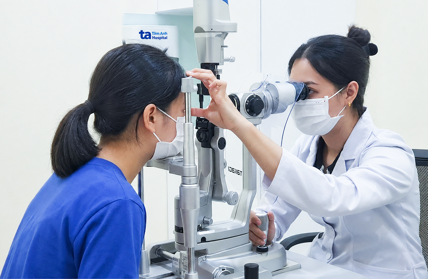 |
Dr. Nhi uses a slit-lamp microscope to examine the child's fundus. *Photo: Tam Anh General Hospital* |
Retinal tears can result from various factors, including trauma, eye surgery, severe myopia, retinal degeneration, or vitreous detachment. Patients may experience symptoms such as floaters, flashes of light, partial vision loss, or a sensation of a curtain obscuring their vision.
Dr. Nhi advises individuals who have sustained eye injuries to undergo a specialized examination, even if there are no open wounds or their vision appears normal. This is crucial for detecting any potential underlying damage. Additionally, athletes and those working outdoors should wear protective eyewear to minimize the risk of injuries and foreign objects entering their eyes.
Ngoc Kim Tham
*Patient's name has been changed
| Readers can submit ophthalmology questions here for doctors to answer. |