Ms. Ngoc presented with a painless swelling in her cheek, initially diagnosed as a benign parotid gland tumor. One month later, she visited Tam Anh General Hospital Ho Chi Minh City (TP HCM) for an ear, nose, and throat endoscopy combined with an MRI. The MRI revealed a tumor with increased vascularity. Master of Science, Doctor, First Degree Specialist Truong Minh Thinh, from the Ear, Nose, and Throat Center, performed a Doppler ultrasound of the patient's neck lymph gland tumor, noting a lesion in the superficial lobe of the left parotid gland, with no neck lymphadenopathy.
Ms. Ngoc then underwent tumor removal surgery and pathological analysis. Professor, Doctor of Science, Doctor Tran Phan Chung Thuy, Director of the Ear, Nose, and Throat Center and head of the surgical team, performed the parotid gland tumor resection on Ms. Ngoc using an AI-powered microsurgical microscope. The procedure involved dissecting the sternocleidomastoid muscle and separating the facial nerve (cranial nerve VII) branch by branch from the tumor and the superficial lobe of the parotid gland to prevent facial paralysis complications. A frozen section biopsy confirmed a benign mixed salivary gland tumor.
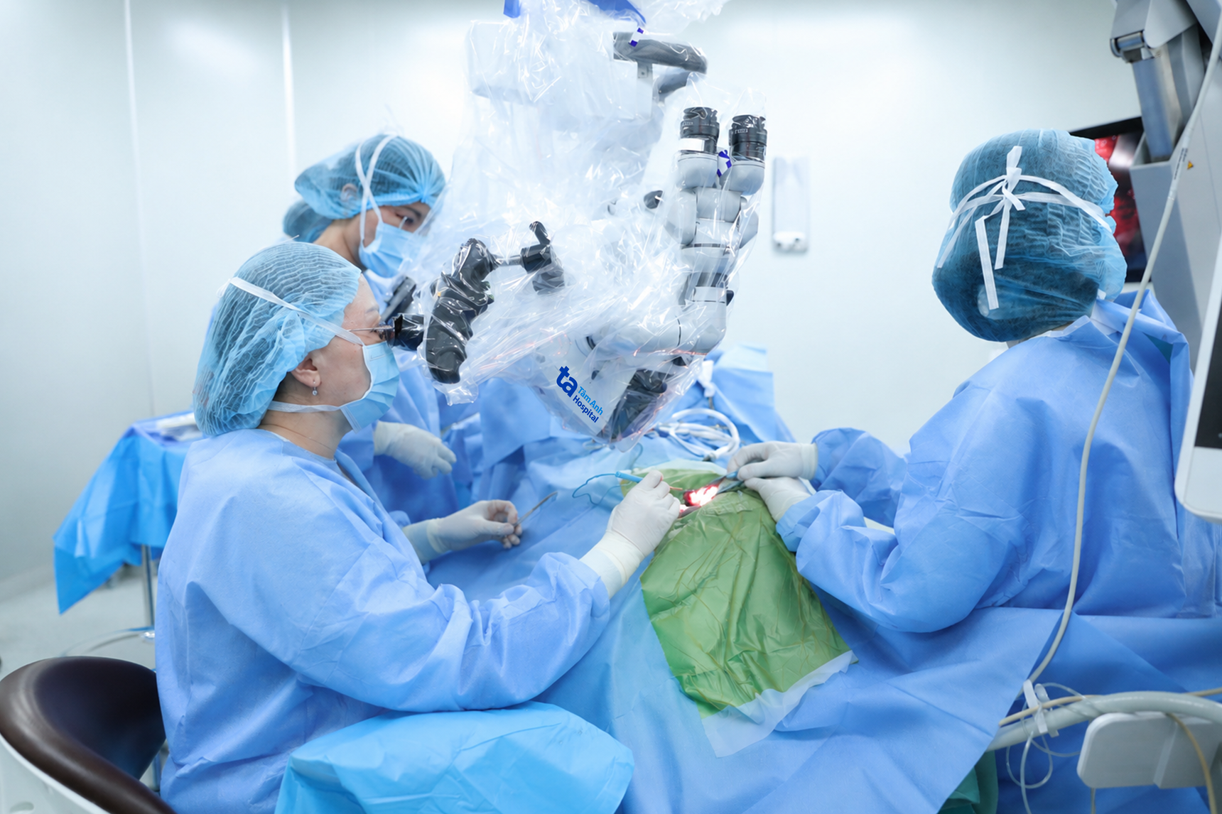 |
Professor Chung Thuy observing through the microsurgical microscope during Ms. Ngoc's tumor removal. Photo: Tam Anh General Hospital
The preceding MRI images showed rapid tumor growth within one month, suggesting potential malignancy. Professor Chung Thuy decided to proceed with a deeper dissection of the superficial lobe of the parotid gland, taking additional samples for pathological analysis. The results from this analysis confirmed carcinoma ex pleomorphic adenoma, minimally invasive type.
According to Professor Chung Thuy, this was an early-stage cancer. The minimally invasive type means that cancer cells had just extended beyond the benign tumor capsule by a very small distance, less than 1,5 mm. With early detection and treatment, and complete removal of malignant cells, the prognosis is excellent: a five-year survival rate of up to 98%. This type carries a low risk of metastasis and recurrence compared to widely invasive forms, where the five-year survival rate is only about 25-65%.
Following surgery, Ms. Ngoc received adjuvant 30-fraction radiation therapy. After completing the treatment, the patient resumed normal activities without any complications of facial paralysis, maintaining regular follow-up appointments.
Professor Chung Thuy explained that pleomorphic adenoma is a benign tumor, and its malignant transformation often progresses silently over many years. The malignant tumor does not develop independently but "hides" within a benign mixed tumor. Malignant cells typically appear in a very small, localized area within the tumor, while most of the surrounding tissue remains benign.
Initially, the condition presents no clear symptoms, existing as a silent, painless, and non-swelling lump for many years. When it turns malignant, the tumor begins to swell rapidly in a short period, which may or may not cause pain. Ms. Ngoc's case, for example, involved no pain, only a rapid increase in tumor size within one month.
Carcinoma ex pleomorphic adenoma (CXPA) is a rare primary salivary gland cancer, typically arising from pre-existing pleomorphic adenomas. It occurs more frequently in individuals 60-80 years old.
Doctors advise patients not to be complacent if a tumor is painless or grows slowly. Instead, patients should undergo regular check-ups so doctors can correctly identify the nature of the tumor, provide appropriate treatment, and prevent disease progression.
Uyen Trinh
*Patient's name has been changed
| Readers can submit questions about ear, nose, and throat conditions here for doctors to answer |