A 12-week prenatal ultrasound at Tam Anh General Hospital TP HCM revealed significant abnormalities in a 27-year-old woman named Minh. The fetus exhibited a disproportionate head-to-abdomen ratio, a small abdomen, and an abnormally enlarged fourth ventricle, a structure containing cerebrospinal fluid in the cerebellum.
Dr. Nguyen Phuong Thao from the Fetal Medicine Center, explained that these findings suggested a risk of early fetal growth restriction or a genetic disorder. An enlarged fourth ventricle signals a structural abnormality of the central nervous system. The fetus was suspected of having triploidy, a condition where it possesses 69 chromosomes instead of the typical 46. Consequently, Minh underwent chorionic villus sampling (CVS) for rapid genetic testing, with results confirming the triploidy diagnosis within 48 hours.
Triploidy is a rare chromosomal abnormality, affecting approximately 1 in 30,000 to 1 in 200,000 pregnancies, according to Dr. Thao. Affected fetuses often show early intrauterine growth restriction, a disproportionate head-to-body size, neural tube defects, limb malformations, and placental abnormalities. Most cases tragically result in miscarriage between 7 and 17 weeks of gestation. For the few who survive to birth, infants typically succumb within the first days of life.
Another case involved Thu, 30, whose 11-week prenatal ultrasound detected a nuchal translucency measuring 5 mm—significantly thicker than the normal measurement of under 3 mm—along with a fetal cystic hygroma in the neck region. Rapid chorionic villus sampling results, available after 48 hours, confirmed the fetus had Edwards syndrome, or trisomy 18. This condition arises from the presence of three copies of chromosome 18 instead of the usual two, with an incidence rate of about 1 in 2,500 pregnancies.
Edwards syndrome severely disrupts fetal development, leading to multiple birth defects. These can affect vital organs such as the heart, brain, musculoskeletal system, digestive system, and urinary system, profoundly impacting the child's life functions.
Dr. Thao noted that fetuses with Edwards syndrome have a high probability of spontaneously ceasing development. For those born with the condition, there is approximately a 50% chance of surviving more than one week, and only about 5-10% live past one year.
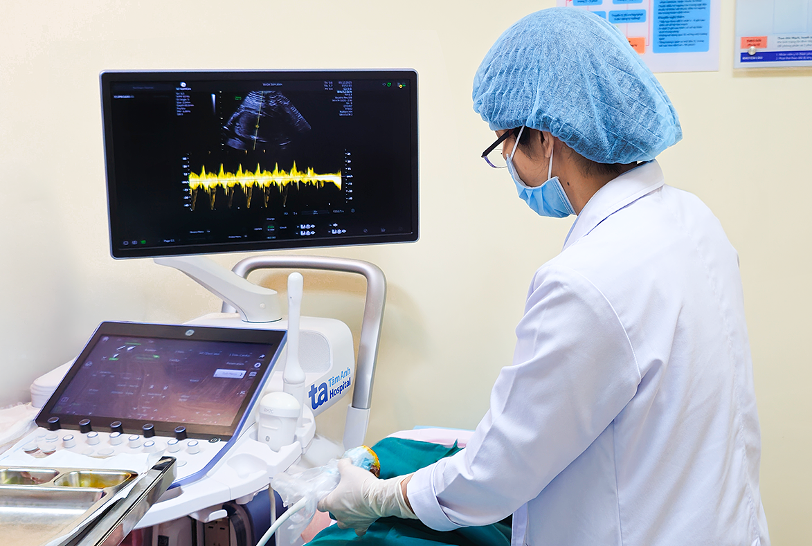 |
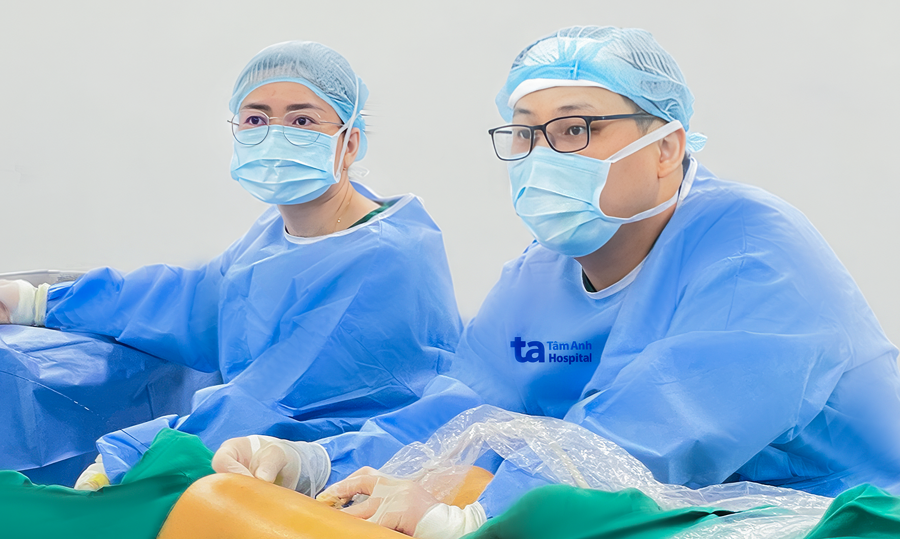
Dr. Thao prepares to collect a chorionic villus sample from Minh with ultrasound guidance. Photo: Tam Anh General Hospital |
Dr. Thao highlighted that many severe fetal malformations can be detected early in the first trimester. Based on specific ultrasound abnormalities, doctors recommend chorionic villus sampling for rapid testing, known as QF-PCR. This test accurately diagnoses numerical abnormalities in five chromosome pairs: 13, 18, 21, X, Y, as well as triploidy, with results typically available in two to three days.
Chorionic villus sampling is a diagnostic procedure performed between week 11 and week 14 of pregnancy. During the procedure, doctors collect a small tissue sample from the placenta. The placenta, which originates from the embryo, contains cells with a genetic code nearly identical to that of the fetus, making it suitable for genetic analysis.
If the rapid test results show no abnormalities, the chorionic villus sample undergoes comprehensive chromosomal analysis. This includes screening for microduplications, microdeletions, and gene mutations. Pregnant women generally receive these full results after about three weeks, though some specialized tests may require a longer turnaround time.
The primary goal of early pregnancy screening is to identify abnormalities. This enables families to receive comprehensive counseling and make informed decisions regarding their pregnancy. Therefore, pregnant women are advised to adhere to their prenatal appointment schedule for timely screening of any potential malformations.
Ngoc Chau
* Patient names have been changed
| Readers can submit questions about pregnancy and childbirth here for a doctor's response |