Results from an MRI and digital subtraction angiography (DSA) at Tam Anh General Hospital Ho Chi Minh City revealed that Lanh had a left dural carotid-cavernous fistula, specifically an indirect carotid-cavernous fistula.
Dr. Nguyen Trung Duc, from the Center for Diagnostic Imaging and Interventional Radiology, explained that dural arterial branches had formed an abnormal connection with the cavernous sinus. This caused arterial blood to flow directly into the venous system, significantly increasing orbital venous pressure. The resulting symptoms included eye redness, conjunctival edema, eye protrusion, glaucoma, oculomotor dysfunction, and reduced vision.
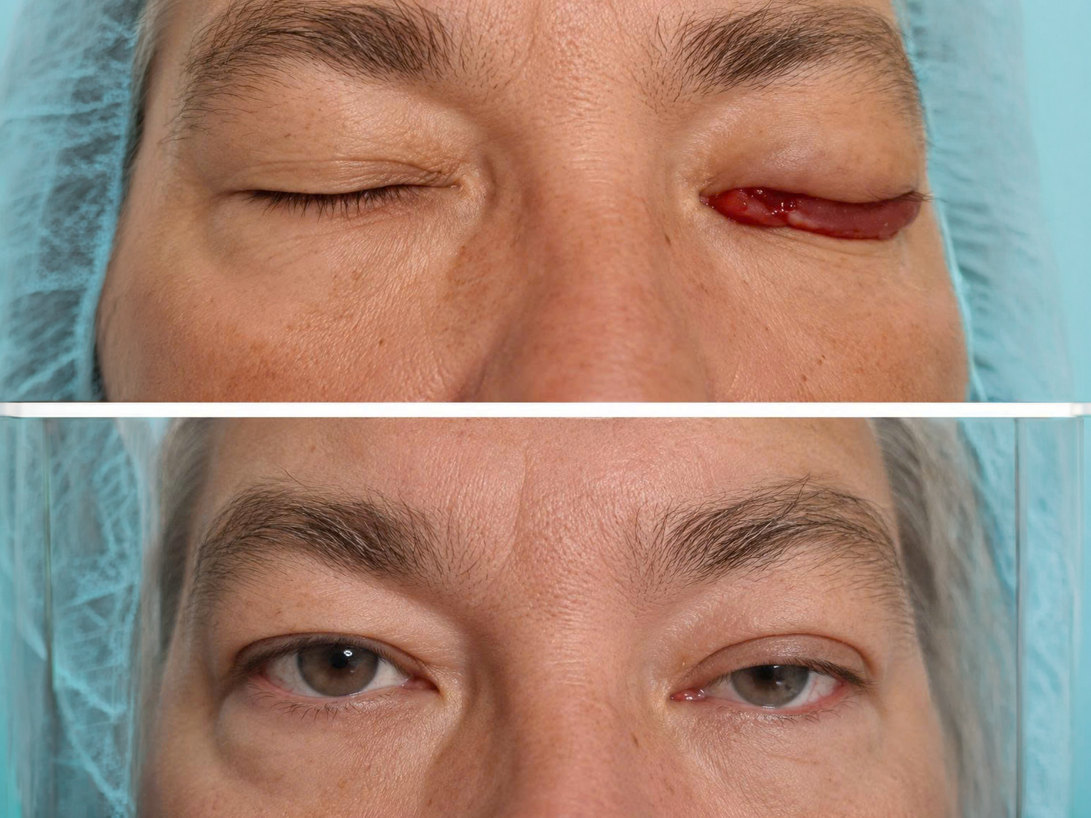 |
Lanh’s left eye before (top) and after (bottom) the intervention. *Tam Anh General Hospital* |
According to Dr. Duc, initial symptoms of this condition often resemble common eye diseases. Consequently, patients are frequently treated for conditions like conjunctivitis, orbital disease, or other ophthalmic disorders before the underlying vascular cause is identified.
Following a multidisciplinary consultation, the medical team opted to treat Lanh with endovascular embolization via a venous approach, guided by digital subtraction angiography (DSA). This technique allows access and treatment of the lesion from within the blood vessel, eliminating the need for open skull base surgery.
Dr. Duc noted that dural carotid-cavernous fistulas are located deep within the skull base, near many blood vessels and nerves that control eye function. For cases presenting with vision loss, glaucoma, oculomotor dysfunction, or dangerous venous drainage pathways identified on DSA, endovascular intervention becomes the preferred treatment.
The medical team guided a microcatheter to the fistula site via the venous route. They then used metallic coils and embolic agents to seal the abnormal connection, effectively closing the fistula.
After two hours of intervention, post-procedure DSA images confirmed the absence of any fistula flow. Lanh reported reduced headaches, and his left eye showed less redness, decreased swelling, and diminished protrusion. Following three days of monitoring, he was discharged in stable condition.
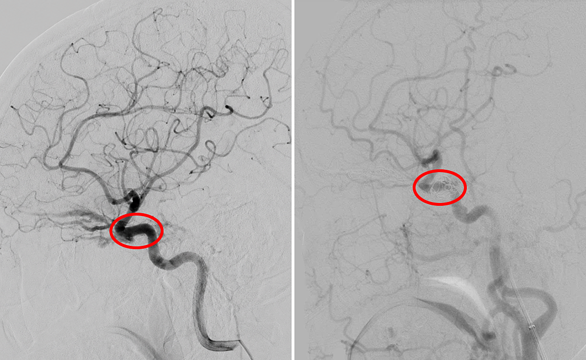 |
The dural carotid-cavernous fistula site in Lanh’s brain before (left) and after (right) vascular intervention. *Tam Anh General Hospital* |
Dr. Duc advises individuals experiencing persistent eye redness, eye protrusion, blurred vision, or headaches that do not respond to conventional treatment to seek further evaluation for underlying neurological and vascular causes.
Nhu Ngoc
*Patient's name has been changed.
| Readers can submit questions about ophthalmic diseases here for doctors to answer. |