An ultrasound at the Fetal Medicine Center, Tam Anh General Hospital, Ho Chi Minh City, revealed that the fetus had multicystic dysplastic kidney on the left side, with dilation of the left renal pelvis and ureter, and a cyst at the distal end of the ureter. Multicystic dysplastic kidney is a congenital anomaly where one or both kidneys form fluid-filled cysts of varying sizes, resembling a cluster of grapes, leading to impaired kidney function.
Master of Science, Doctor Ngo Thi Binh Lua, from the Obstetrics and Gynecology Center, stated that most cases of multicystic dysplastic kidney are isolated defects. Some cases may be accompanied by other structural abnormalities, such as those affecting the digestive, cardiovascular, or nervous systems, or may be linked to chromosomal abnormalities or gene mutations. The 29-year-old expectant mother was advised by doctors to undergo amniocentesis to determine if this was an isolated defect or due to a genetic abnormality, but the couple declined for personal reasons.
Doctors assessed that the fluid-filled cyst at the end of the ureter, connecting to the bladder, likely caused retrograde obstruction. This obstruction led to urine retention, dilating the ureter and renal pelvis, and ultimately causing multicystic dysplastic kidney. The retrograde obstruction caused the left kidney to enlarge, potentially compressing adjacent organs. The fetus faced risks of kidney failure and loss of function in the left kidney. The right kidney remained unaffected, with a prognosis for stronger development to assume the function of the left kidney.
After birth, the child may experience urinary tract infections or vesicoureteral reflux. Depending on the case, the patient might require surgery to remove the left kidney or treatment for kidney failure in adulthood. If the remaining kidney also exhibits abnormalities and impaired function, the patient would need a kidney transplant.
The expectant mother underwent close monitoring of amniotic fluid volume and the development of the right kidney. A comprehensive fetal echocardiogram showed no abnormalities. At 28 weeks of gestation, an ultrasound revealed a partial duplex kidney, meaning the left kidney had two separate renal pelves with two distinct ureters. These two ureters then merged into a single tube before entering the bladder. This condition can lead to further dilation of the urinary system or an increased risk of urine reflux.
At 33 weeks of gestation, the fetus was small for its gestational age, raising concerns about intrauterine growth restriction and fetal demise. Doctors provided counseling on appropriate nutrition and exercise, guiding the expectant mother to monitor fetal movements.
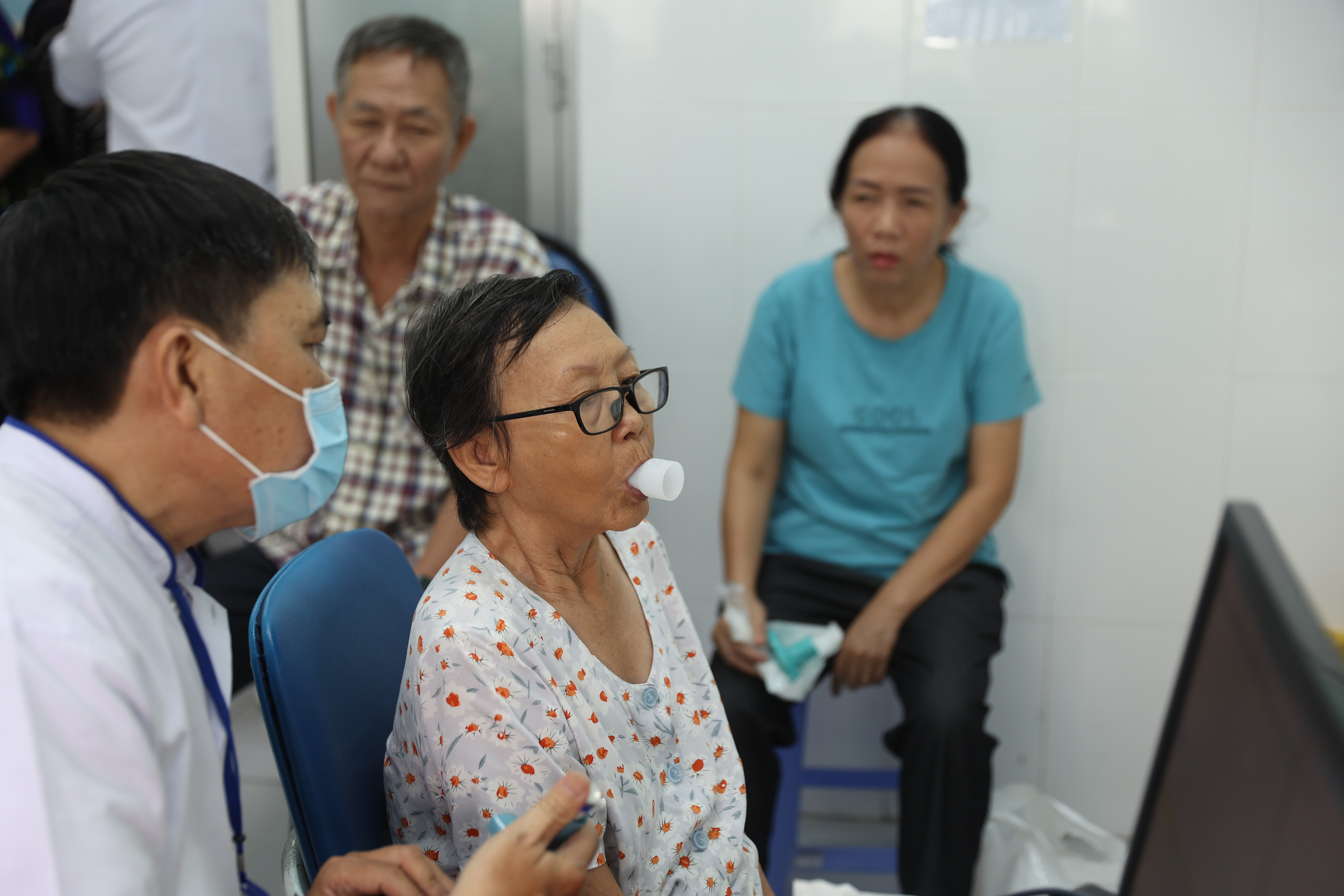
The baby boy was born at 39 weeks of gestation, weighing approximately 2,7 kg, with no immediate postnatal health abnormalities. He requires regular check-ups with a pediatric nephrology and urology specialist for timely intervention and to prevent complications.
 |
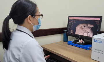
Doctor Lua (right) and her team performed a cesarean section for the expectant mother. Photo: Tam Anh General Hospital |
Doctor Lua cited research indicating that multicystic dysplastic kidney occurs in 1/1,000 to 5,000 live births, with approximately 75-80% being isolated abnormalities. Typically, only one kidney is affected, while the other kidney remains healthy and grows larger to perform the function of both. In rare instances, both kidneys are affected, which can lead to underdeveloped fetal lungs (pulmonary hypoplasia), oligohydramnios, and a high risk of death before or after birth.
For unilateral multicystic dysplastic kidney, the abnormal kidney tends to shrink and disappear over time. After birth, children need regular ultrasound monitoring every six months or one year. This allows doctors to assess the degree of atrophy and ensure no tumors form in the kidney. The healthy kidney is also screened for obstruction or reflux. Most infants with multicystic dysplastic kidney remain healthy if the remaining kidney functions well.
Doctor Lua advises expectant mothers who discover any abnormalities during routine prenatal check-ups to seek care at specialized obstetrics units. This ensures individualized pregnancy management and preparation for appropriate postnatal treatment plans, safeguarding both mother and baby.
Ngoc Chau
| Readers can submit questions about obstetrics and gynecology here for doctors to answer. |