On 26/3, representatives from K hospital announced that its Quan Su facility (campus 1) has been equipped with eight new endoscopy systems. A highlight is the EVIS X1 system for digestive and bronchial endoscopy, which features virtual chromoendoscopy and optical image magnification up to 150 times, aiding in the precise diagnosis and intervention of cancerous lesions.
These machines were acquired through a 790 billion VND investment from the state budget and 300 billion VND in non-refundable aid sponsored by the Japanese government. On 10/3, the hospital also began operating two new-generation accelerator radiotherapy systems, which help doctors shorten treatment time per case from 10 minutes to two or three minutes, significantly reducing prolonged patient wait times.
Doctor Bui Anh Tuyet, Head of the Department of Endoscopy and Functional Exploration, stated that esophageal cancer is among the 15 most common cancers, with 3,686 new diagnoses and 3,470 deaths annually. Most esophageal cancer patients are diagnosed at a late stage, presenting with clear symptoms such as difficulty swallowing, progressive choking, and hoarseness due to tumor invasion into surrounding tissues. At this advanced stage, surgery is often not possible, and concurrent chemoradiotherapy is usually indicated, though the prognosis is not favorable.
"Early diagnosis plays a decisive role in the treatment and prognosis of esophageal cancer, significantly increasing treatment effectiveness and reducing costs," Doctor Tuyet stated. According to multiple studies in Japan, the five-year survival rate for patients diagnosed early can exceed 90%.
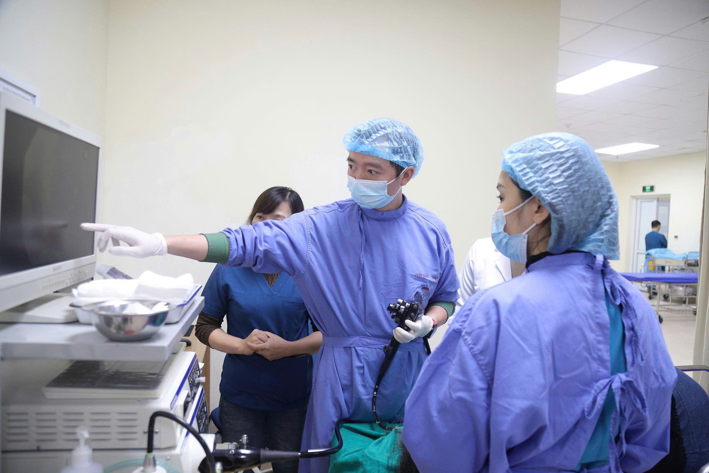 |
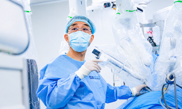
Doctors perform endoscopy for early screening and diagnosis of digestive tract cancer in patients. *Photo: Ha Tran*
Previously, diagnosing esophageal cancer primarily relied on endoscopy to identify lesions and biopsies for histopathological examination. However, conventional white-light flexible endoscopy often found it challenging to detect early lesions in the esophagus because they are small and have a color similar to the surrounding mucosa. Now, the new endoscopy system features light-based chromoendoscopy and over 150x magnification, which clearly analyzes the microstructure and microvessels of lesions, making diagnosis and treatment easier for doctors.
"The purpose of magnified endoscopy is to facilitate early detection and differentiate common digestive tract cancerous lesions found in the stomach, esophagus, and colorectum," Doctor Tuyet explained. Applying advanced image processing technologies for cancer diagnosis and classification is not merely for early screening and detection; the hospital's primary goal is to treat cancerous lesions at a very early stage through endoscopic intervention.
For example, a 63-year-old male patient with a long history of smoking was scheduled for an upper gastrointestinal endoscopy as part of a routine health check-up. During the procedure, doctors discovered a flat, brownish lesion approximately 15 mm in size. The patient was treated with endoscopic submucosal dissection (ESD), a minimally invasive intervention that limits invasiveness, reduces costs, and improves the patient's survival time and quality of life. Following the ESD procedure, histopathological results confirmed pT1a esophageal squamous cell carcinoma with negative resection margins. The patient recovered well and was discharged after two days.
Previously, esophageal cancer was commonly observed in individuals over the age of 50. However, this disease is now trending towards an increase and affecting younger demographics. Therefore, doctors recommend that everyone undergo regular health check-ups and early screening for timely treatment.
Le Nga