A contrast-enhanced abdominal CT scan of Mr. Thoi at Tam Anh General Hospital in Ho Chi Minh City revealed multiple pseudoaneurysms in various locations, including the hepatic artery, stomach region, kidneys, and lumbar area. Notably, a large pseudoaneurysm measuring 40x40x65 mm was found in the hepatic hilum, originating from a branch of the right hepatic artery. This mass was compressing adjacent structures, causing impaired perfusion of the liver parenchyma and compressing the right renal vein.
Master, Doctor, Specialist Level I Pho Thien Phuoc from the Emergency Department explained that a pseudoaneurysm occurs when a vessel wall tears or is damaged. Blood escapes and is contained by surrounding tissue, forming an abnormal blood sac. Without the protection of the vessel wall, pseudoaneurysms are highly susceptible to rupture, especially when large or located in critical areas such as the liver, kidneys, or stomach.
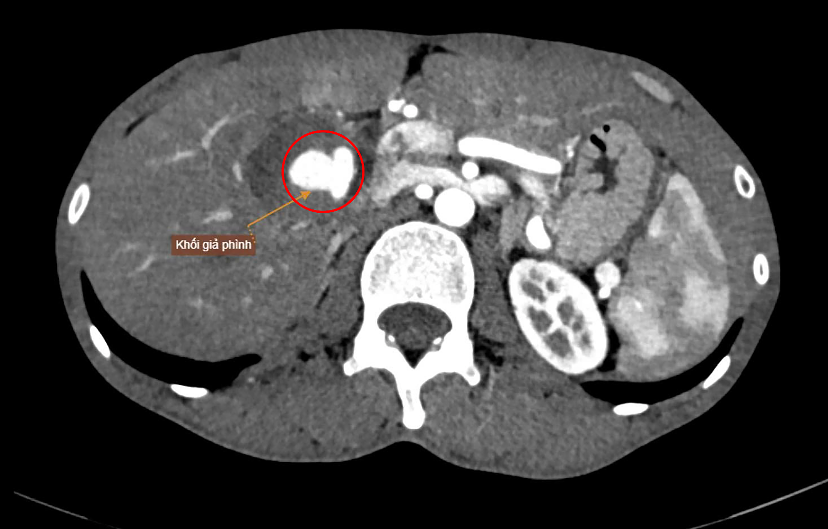 |
A large pseudoaneurysm with rupture risk shown on a CT scan. *Photo: Tam Anh General Hospital* |
Following a consultation, doctors opted for endovascular intervention for the patient, considering it less risky than open abdominal surgery. Guided by a digital subtraction angiography (DSA) system, doctors threaded microcatheters through the arterial system to precisely access the pseudoaneurysm in the liver. The team used metal coils combined with specialized biological glue for selective embolization, effectively sealing the pseudoaneurysm and preventing blood flow into the damaged area, while preserving healthy hepatic branches.
Doctor, Specialist Level I Nguyen Trung Duc from the Endovascular Intervention Unit stated that the patient's condition, characterized by multiple scattered pseudoaneurysms, weak vessel walls, accompanying hematoma, and anatomical deformation of the hepatic hilum, made the intervention challenging. Post-procedure imaging confirmed that the pseudoaneurysm in the liver was completely occluded.
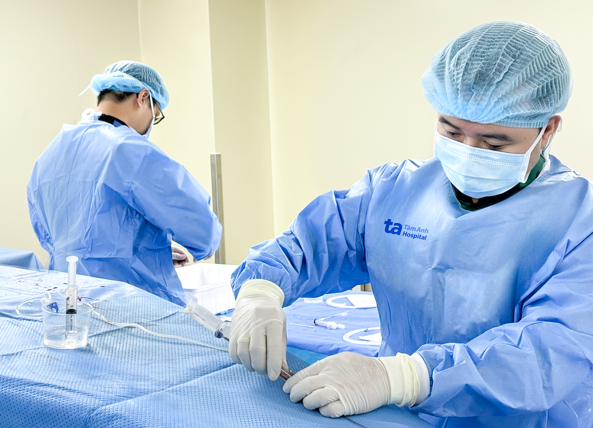 |
Doctor Duc (right) and his team perform an intervention to seal Mr. Thoi's ruptured pseudoaneurysm. *Photo: Tam Anh General Hospital* |
After the intervention, Mr. Thoi's abdominal pain subsided, and his liver perfusion improved. A follow-up abdominal Doppler ultrasound after discharge confirmed the effective intervention on the hepatic artery pseudoaneurysm, with no blood flow detected into the aneurysm sac.
Mr. Thoi's presentation of multifocal pseudoaneurysms led Doctor Duc to suspect a connection to connective tissue abnormalities or primary vessel wall weakness. Consequently, the remaining pseudoaneurysms in his stomach, kidneys, and lumbar area still pose a risk of complications. The patient requires long-term monitoring to assess complication risks and consider preventive interventions like embolization or appropriate medical treatment to prevent rupture.
Doctor Duc advises anyone experiencing unusual symptoms, such as persistent abdominal pain, to seek medical examination for a diagnosis.
Nhat Thanh
*Patient's name has been changed*
| Readers can ask questions about digestive diseases here for doctors to answer |