Doctor Chu Thi Ha, Head of Respiratory Unit at Tam Anh General Clinic, District 7, examined Kien's lungs, noting wheezing in both bronchi from narrowed airways. Laboratory results showed D-Dimer, a blood clot marker, slightly elevated above 500 ng/mL. Doctors diagnosed him with acute exacerbated chronic obstructive pulmonary disease (COPD) and other conditions: hypertension, dyslipidemia, and sleep disorders. He received treatment with antibiotics, anti-inflammatories, mucolytics, and bronchodilators, with a follow-up after three days.
He returned for his follow-up six days late, still coughing; his shortness of breath improved, but he felt fatigued. As his D-Dimer index continued to rise, doctors ordered a triple rule out computed tomography (CT) scan with contrast injection, ultra-thin slices, and multi-phase reconstruction. This diagnostic technique simultaneously evaluates three dangerous conditions with similar symptoms: pulmonary embolism, coronary artery disease, and aortic dissection.
Results revealed Kien also had atherosclerotic plaques causing 50-60% narrowing of the left anterior descending artery, atherosclerosis along the aortic wall, and complete occlusion by thrombi of branches A1, A2, and the distal segment of an A3 branch of the right pulmonary artery. Doctors diagnosed him with pulmonary embolism, complicated by acute exacerbated COPD with superinfection and various cardiovascular and metabolic diseases. Untreated pulmonary embolism disrupts blood flow to the lungs, causing systemic oxygen deprivation, hypotension, arrhythmias, and cardiac arrest.
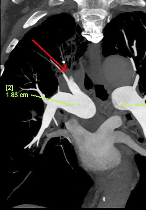 |
Chest CT scan revealing thrombi completely filling the lumen of multiple branches of the right pulmonary artery (red arrows). *Photo: Tam Anh General Hospital* |
Doctors prescribed Kien injectable anticoagulants for eight days, then switched to oral anticoagulants, combined with antibiotics, coronary vasodilators, antiplatelet agents, and lipid-lowering medications. After 12 days of intensive treatment, his shortness of breath and chest pain decreased, his health stabilized, and he was discharged.
Many factors can lead to blood clot formation in the lungs, as seen in Kien's case. Underlying conditions like COPD alter intrathoracic pressure, affecting the heart's pumping function. Patients with vascular wall damage from atherosclerosis, surgery, stent placement, immobility, or prolonged sitting can experience blood flow stagnation. Cancer, autoimmune diseases, and lower limb varicose veins also increase embolism risk. "This is a common cause of cardiovascular death, ranking only after myocardial infarction and stroke," said Doctor Ha.
 |
Doctor Ha consults with Kien during his follow-up appointment. *Photo: Tam Anh General Hospital* |
Pulmonary embolism symptoms vary, including shortness of breath during exertion or at rest, chest pain, fainting, hemoptysis, dizziness, sweating, fever, and cyanosis. Some cases progress silently, appearing suddenly and severely.
Doctor Ha advises elderly individuals with underlying conditions to strictly adhere to treatment regimens, take prescribed medications, and attend follow-up appointments on schedule. When acute exacerbations occur with chronic illness, patients should not arbitrarily stop medication or delay follow-up visits.
Quyen Phan
*Patient's name has been changed*
| Readers can submit questions about respiratory health here for doctors to answer |