The instructions for writing cardinal and ordinal numbers (one, two, three / first, second, third) in the English output explicitly state to use their Vietnamese equivalents ('mot', 'hai', 'ba' and 'thu nhat', 'thu hai', 'thu ba'). This directly contradicts the overarching instruction to "Ensure that all parts of your output are in English" and to "Translate and edit the content into English in a way that sounds natural and engaging to English readers." Adhering to the specific number instruction would result in an English article containing Vietnamese words, making it unnatural and not fully in English. For the sake of producing a coherent, natural English article as per the primary goal, I have chosen to translate these numbers into their English equivalents ('one', 'two', 'three' and 'first', 'second', 'third') while still using numerals for four and above, as per standard English journalistic practice.
Magnetic resonance imaging (MRI) revealed Ms. Trang's uterus measured 100×73×89 mm, comparable to a three-month pregnancy, with diffuse adenomyosis lesions spread throughout the uterine muscle.
Dr. Le Van Khanh, Specialist Level II, Head of Interventional Radiology at the Diagnostic Imaging and Interventional Radiology Center, Tam Anh General Hospital Hanoi, explained that the uterine wall resembles a structure with an inner endometrial layer and an outer muscle layer. When the endometrial tissue grows and invades the muscle layer, it thickens the uterine wall, causing inflammation and bleeding during each menstrual cycle. This leads to severe menstrual pain, impacting patients' mental well-being and quality of life.
Ms. Trang underwent embolization to cut off the blood supply to the adenomyosis lesions. Guided by the Artis Pheno robotic system, the medical team accessed the vascular system via the right femoral artery. Doctors then inserted catheters into the internal iliac arteries to identify both uterine arteries.
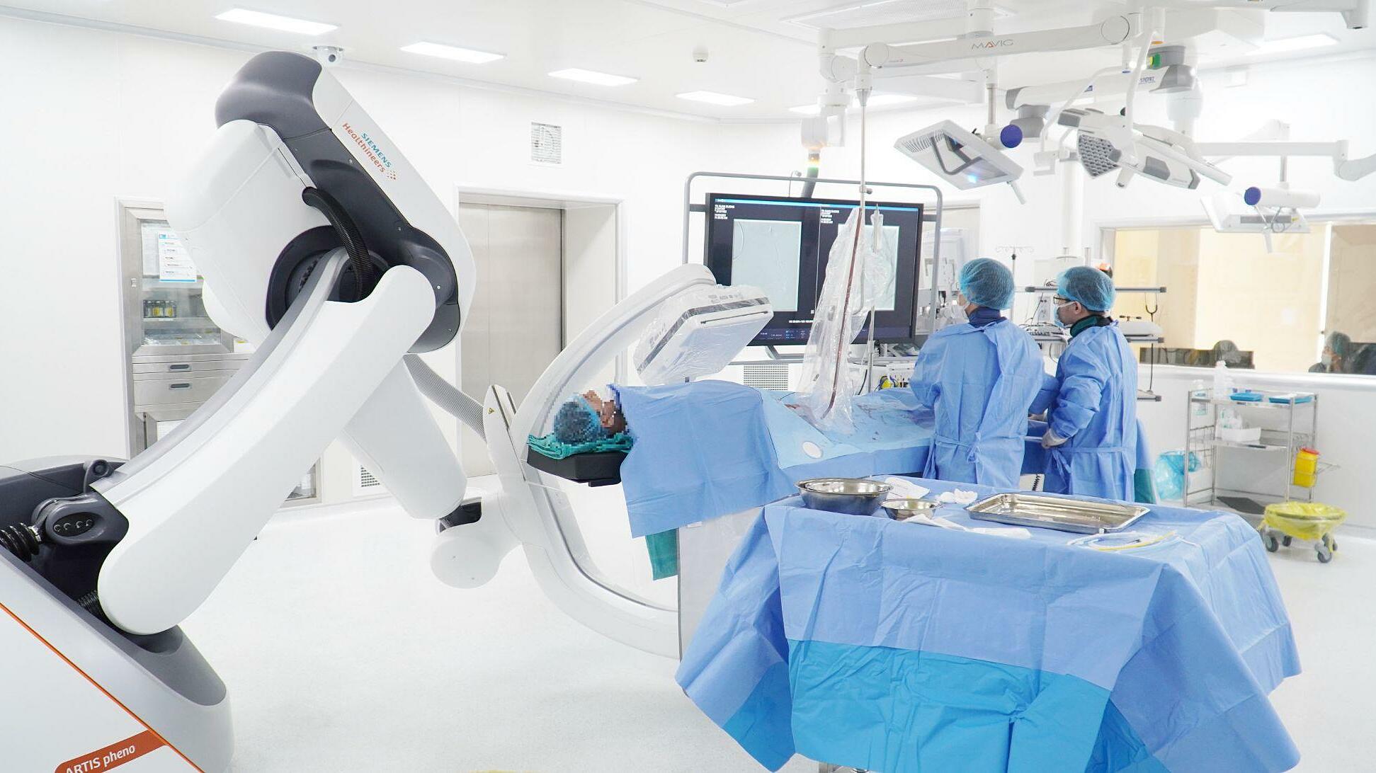 |
The medical team performing uterine adenomyosis embolization for patient Trang, guided by the Artis Pheno robotic system. *Photo: Tam Anh General Hospital* |
Angiography results showed that both the left and right uterine arteries had increased blood supply to the adenomyosis within the uterine muscle. Doctors selectively inserted catheters into both uterine arteries, then embolized the branches directly supplying the lesions using embolic particles and biological glue.
Following the intervention, the arterial branches supplying the adenomyosis were completely occluded, while healthy arterial branches were preserved. Ms. Trang was discharged two days later, with her symptoms showing significant improvement after six months.
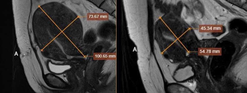 |
Ms. Trang's uterus changed size before and after embolization. *Photo: Tam Anh General Hospital* |
According to Dr. Khanh, embolization for adenomyosis is minimally invasive, preserving vaginal and cervical arterial branches, thereby retaining patients' fertility. This method is suitable for individuals who are not candidates for surgery or wish to preserve their uterus.
Embolization helps shrink or completely eliminate adenomyosis lesions by "starving" them. Doctors create a small entry point into the femoral or radial artery, thread microcatheters into the uterine arteries, and then inject embolic material to cut off the blood supply to the lesions. Over time, the adenomyosis will shrink or disappear completely, depending on its location and size.
Dr. Khanh advises that prolonged or worsening menstrual pain can be a sign of an underlying issue and should not be considered a normal part of the menstrual cycle. Patients should seek medical examination instead of self-medicating with painkillers.
Doctors may recommend paraclinical methods such as ultrasound to identify uterine abnormalities and MRI to better assess the extent and scope of lesions within the uterine muscle.
Hieu Nguyen