Mr. Phuong has a history of hypertension, diabetes, and dyslipidemia, all managed with medication. For the past month, he experienced occasional severe chest pain, prompting him to visit Tam Anh General Hospital, TP HCM. Master of Science, Doctor, Level II Specialist Ho Anh Tuan from the Interventional Cardiology Center, reported that the patient's coronary artery system was narrowed. Specifically, the middle segment of the right coronary artery showed 50-60% narrowing, the middle segment of the left anterior descending artery was 60-70% narrowed, and the circumflex artery had 70-80% narrowing at its origin. Plaque and calcification caused the narrowing in all three branches.
Professor, Doctor Vo Thanh Nhan, Director of the Interventional Cardiology Center, explained that stent placement is typically used for narrowing above 70%, or 50-70% in high-risk areas like the left main coronary artery or proximal left anterior descending artery, especially when accompanied by symptoms such as exertional angina. Mr. Phuong's degree of narrowing could potentially be managed medically. However, since all three of his coronary artery branches were affected by plaque and calcification, it was necessary to assess the nature of the plaque to determine the risk of myocardial infarction.
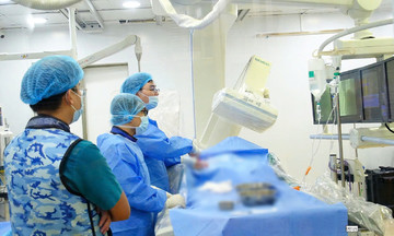
Doctors utilized optical coherence tomography (OCT), a super-high-resolution imaging technique 10 times more precise than intravascular ultrasound (IVUS), to observe the micro-structure of the vessel wall in detail. OCT revealed widespread, mixed plaques with a high risk of rupture, which conventional coronary angiography might miss. "If stent placement for the narrowed coronary artery branch is not performed early, the plaque could rupture, causing acute myocardial infarction," Professor Nhan stated.
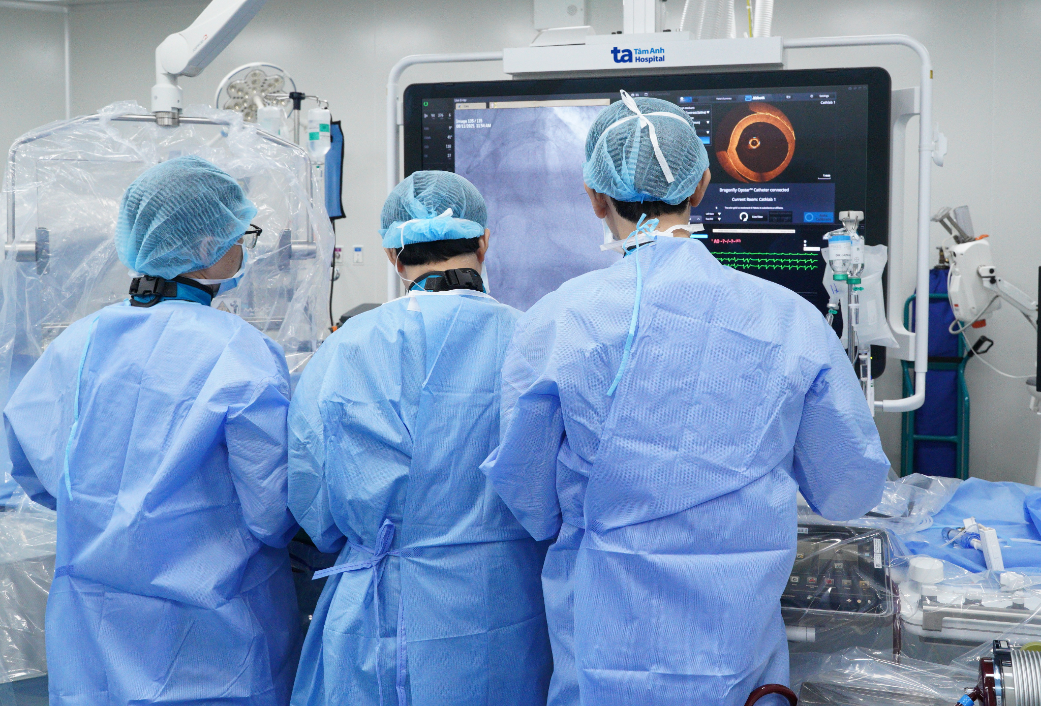 |
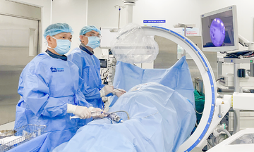
Doctors perform stent placement for the patient with the support of OCT technology. Photo: Ngoc Ha |
Doctors identified the left anterior descending artery as having a high risk of occlusion due to easily rupturable plaque and decided to place a stent in this branch. Based on the OCT images, the team observed a thin cap fibro-atheroma plaque, which is prone to rupture. They precisely measured the vessel size and lesion length to select an appropriate stent. Following placement, the team confirmed the stent was fully expanded and well-apposed to the vessel wall, with no dissection at either end, minimizing the risk of acute post-stent occlusion.
After the intervention, Mr. Phuong was alert and free of chest pain. He was discharged after 3 days, with the risk of myocardial infarction significantly reduced. Post-stent placement, doctors advised the patient to adhere to medical treatment, maintain a healthy lifestyle (quit smoking, limit alcohol, increase physical activity, avoid stress), manage underlying conditions, and attend follow-up appointments.
Thu Ha
* Patient's name has been changed
| Readers can submit questions about obstetrics and gynecology here for doctors to answer. |