According to Master of Science, Doctor Dang Quy Loi from the Obstetrics and Gynecology Unit at Tam Anh Cau Giay General Clinic, an echogenic intracardiac focus (EIF) is a small bright spot appearing in the fetal heart chamber, with echogenicity similar to bone on ultrasound images. This finding is related to a mild calcium deposit in the papillary muscle or chordae tendineae within the heart; it is not a structural abnormality and does not affect the heart's contractile function.
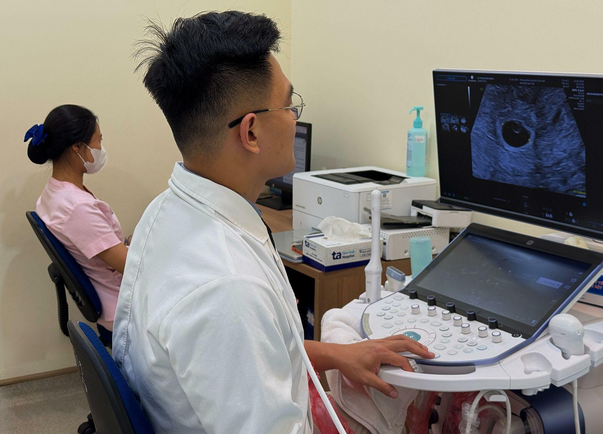 |
A doctor performs an ultrasound to check fetal development. Photo: Tam Anh General Hospital
An EIF in the fetal heart typically measures 1-4 mm, most commonly 2-3 mm, and is observed in approximately 4-5% of pregnancies during second-trimester ultrasounds. Among these cases, about 86% are in the left ventricle, 3% in the right ventricle, and 10% appear on both sides of the heart. The size of the EIF is not a factor in assessing its severity; the more important consideration is whether other abnormalities are present.
This finding is usually detected during the fetal morphology ultrasound performed between 18 and 22 weeks of gestation. An EIF can appear in the first trimester, but the fetal heart is still small, making detection difficult.
An EIF does not cause heart defects, impair heart function, or affect fetal development. Many studies tracking children after birth show that most children with an isolated EIF have completely normal hearts, requiring no treatment or cardiovascular monitoring solely due to this finding.
In obstetrics, EIF was once considered a marker for the risk of chromosomal abnormalities. However, this does not mean the fetus definitely has a condition; it merely suggests that doctors should investigate further if other accompanying abnormalities are present.
Previously, before the advancement of prenatal screening methods, many doctors considered the presence of an EIF on ultrasound as a factor increasing the likelihood of the fetus having Down syndrome (trisomy 21). Today, more accurate screening tests like the double test, triple test, and especially non-invasive prenatal testing (NIPT), are available.
If a fetus has only one EIF in the heart, no other abnormalities are detected on ultrasound, and prenatal screening results indicate a low-risk group, no further invasive tests are needed. Pregnant women simply continue routine prenatal care.
If the fetus has other indicators such as increased nuchal translucency, kidney pelvis dilation, a short nasal bone, heart abnormalities, or if the mother is in a high-risk group (advanced maternal age, family history), the doctor may recommend additional testing.
In many cases, an EIF will fade or no longer be visible in the third trimester of pregnancy. However, some fetuses may still have it until late pregnancy or after birth. This does not impact the child's health.
Doctor Loi advises expectant mothers not to be overly concerned if their fetus has an EIF in the heart. The crucial step is to evaluate the ultrasound results within the context of the entire pregnancy, including gestational age, morphological ultrasound findings, prenatal screening results, and individual risk factors for each pregnant woman.
Thuy Hanh
| Readers can submit questions about pregnancy and childbirth here for doctors to answer. |