A macular hole is damage occurring in the macula, a critical part of the retina responsible for central vision. This area enables activities such as reading, recognizing faces, watching tivi, and perceiving fine details. The condition is common in older adults, typically developing as the vitreous gel shrinks due to aging, pulling on the retina and gradually forming a hole in the macula.
While not life-threatening, a macular hole poses a significant risk to vision. It can cause noticeable central vision loss, blurred vision, distorted images, or a dark spot in the center of one's visual field. Without treatment, the condition typically worsens, leading to prolonged vision impairment and impacting daily activities.
Currently, the effective treatment is often a vitrectomy. This surgical procedure involves removing the vitreous gel, peeling the internal limiting membrane, and injecting intraocular gas to help the macular hole close. Modern techniques achieve high success rates, especially when the condition is detected and treated early.
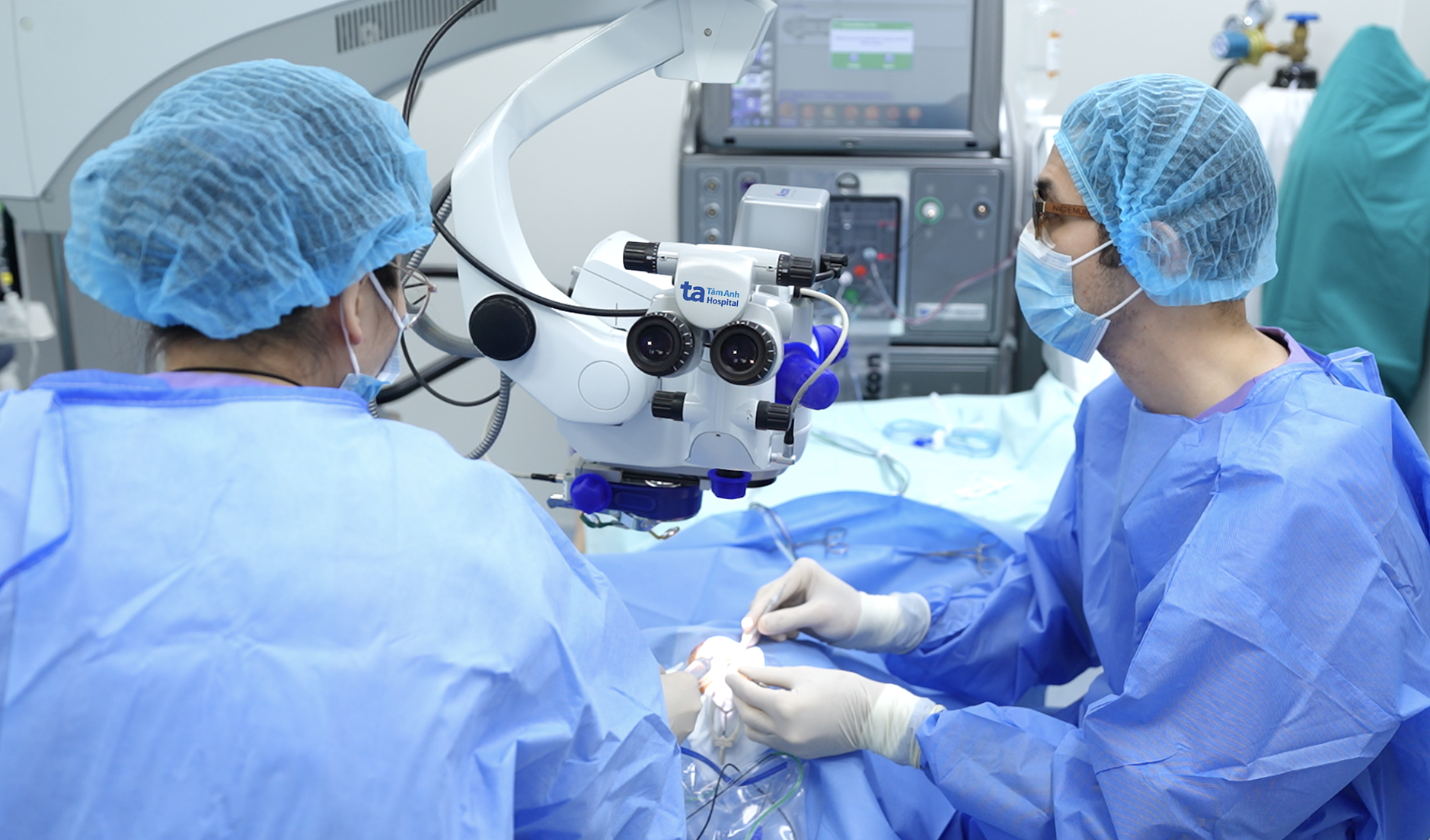 |
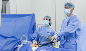
A doctor performs a vitrectomy. Illustration: Tam Anh General Hospital |
Beyond surgery, for some very early-stage, small macular holes, doctors may consider intraocular injections. These injections aim to assist in detaching the vitreous from the macula, reducing traction, and facilitating the hole's natural closure. However, their effectiveness is less consistent than surgery, and they are not widely adopted across all medical facilities. A vitreoretinal specialist must thoroughly evaluate the patient's condition before prescribing this option.
The retina, rich in optic nerves, means damage in this area cannot fully recover. Many macular holes may close well after surgery, but vision often does not return to its previous state because photoreceptor cells in the macula have been damaged. The extent of recovery depends on the hole's size, disease stage, duration of the condition, and any co-existing pathologies such as cataracts or age-related macular degeneration.
Following surgery, some patients must lie face down as instructed. This position helps the gas bubble apply pressure correctly, aiding in better hole closure. Patients also require regular follow-up appointments to monitor progress and check the other eye, as macular holes can develop in both eyes.
Given this, your father should visit a medical facility or hospital with a Vitreoretinal department. There, he can undergo an evaluation using optical coherence tomography (OCT) and receive appropriate treatment advice. Early intervention often improves recovery chances and limits prolonged central vision loss.
Dr. Bui Viet Hung
Head of Vitreoretinal Department,
High-Tech Eye Center
Tam Anh General Hospital
| Readers can submit ophthalmology questions here for doctors to answer |