Answer:
Ultrasound allows doctors to assess fetal development, the condition of the uterus and placenta, and detect early health abnormalities in both mother and baby. During a normal pregnancy, mothers typically undergo several ultrasounds.
The first milestone occurs at 6-8 weeks of pregnancy: As soon as a positive pregnancy test result or a missed period is noted, many women are advised to have an early ultrasound to confirm if the pregnancy is located within the uterus.
At this stage, an ultrasound helps confirm the implantation site, ruling out the risk of an ectopic pregnancy, a dangerous condition if not detected promptly. Doctors also check the number of fetuses, evaluate the gestational sac, yolk sac, and fetal heart activity.
Fetal heart activity can typically be detected around weeks 6-7 of pregnancy. This timing also provides a relatively accurate estimate of gestational age, which serves as a basis for monitoring fetal development in subsequent months.
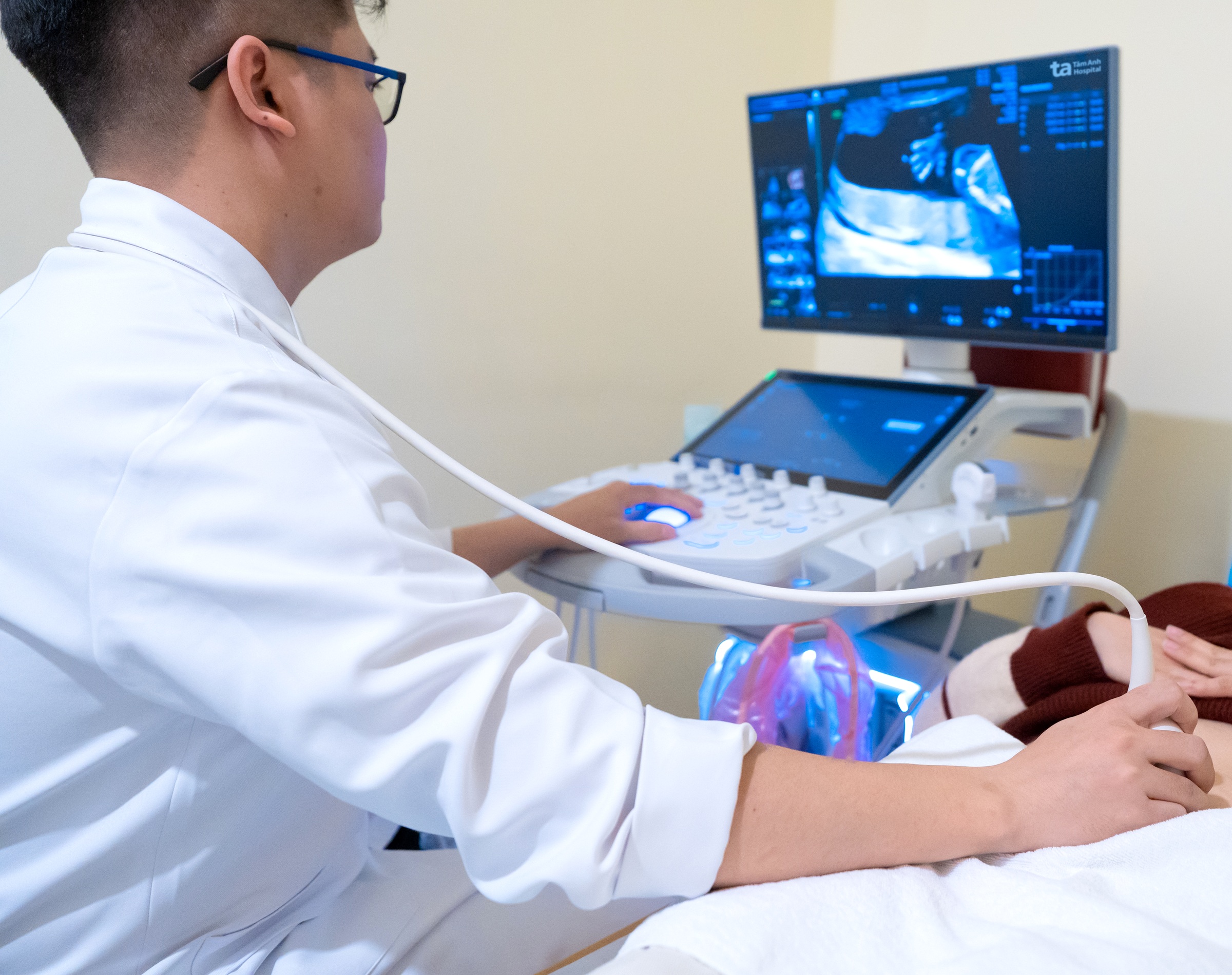 |
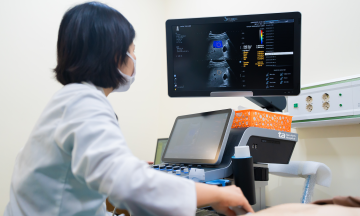
A doctor performs an ultrasound to assess fetal development. Photo: Tam Anh General Hospital
The 11 weeks to 13 weeks and 6 days milestone involves nuchal translucency measurement: This is one of the most critical ultrasounds during the first trimester of pregnancy.
Nuchal translucency is a fluid collection behind the fetal neck. An abnormally increased measurement can indicate a higher risk of chromosomal disorders such as Down, Edwards, or Patau syndromes.
Additionally, an ultrasound at this stage allows doctors to assess the presence of the nasal bone, conduct an early survey for certain morphological abnormalities, and combine findings with prenatal screening tests to calculate the risk of congenital defects.
The 18-22 weeks milestone: When the fetus reaches approximately 18-22 weeks of age, its organs have developed enough for doctors to conduct a detailed anatomical survey. Through a morphological ultrasound, doctors evaluate the fetal brain, skull, face, lips, spine, heart, lungs, stomach, kidneys, bladder, abdominal wall, and limbs.
The primary goal is to detect congenital defects identifiable via ultrasound, such as cleft lip, central nervous system abnormalities, or congenital heart defects. Beyond the fetus, doctors also assess the placental position, cervical length, amniotic fluid volume, and the fetal growth rate.
At 22-24 weeks, a specialized fetal echocardiogram is performed: This examination is often indicated when the pregnant woman has risk factors, such as a family history of congenital heart disease or pre-pregnancy diabetes. A fetal echocardiogram provides a detailed assessment of the heart chambers, heart valves, great vessels, and blood flow within the heart, allowing for early detection of many congenital heart conditions.
The 28-32 weeks milestone: During the third trimester, the ultrasound focus shifts to evaluating the fetal growth rate.
Doctors will measure biometric parameters, including biparietal diameter, head circumference, abdominal circumference, and femur length, to estimate fetal weight. This data helps detect intrauterine growth restriction or a fetus that is too large for its gestational age.
An ultrasound at this stage also helps assess amniotic fluid volume, placental function, and umbilical blood flow in necessary cases. These are crucial factors reflecting the oxygen and nutrient supply to the fetus.
If inappropriate fetal growth or signs of fetal distress are detected, doctors may adjust the monitoring and treatment plan to reduce the risk of complications for both mother and baby.
The 35-37 weeks milestone: The final weeks of pregnancy are when doctors gather essential information for childbirth. An ultrasound helps determine fetal presentation, estimated weight, placental position, amniotic fluid volume, and umbilical cord status. These are important factors for predicting the likelihood of a vaginal birth or considering a cesarean section when necessary.
For example, if the fetus is in breech presentation, the placenta is low-lying, or severe oligohydramnios occurs, doctors will devise an appropriate management plan to limit risks during labor.
Additionally, close monitoring in the final stage helps detect early signs of abnormalities such as growth restriction, placental dysfunction, or the risk of stillbirth.
Diagnostic ultrasound, when performed with proper indication, is a safe method during pregnancy. However, the number of ultrasounds should be determined by a doctor based on the health status of the mother and fetus.
High-risk pregnancies, such as those involving gestational hypertension, gestational diabetes, or multiple pregnancies, require more frequent monitoring compared to normal pregnancies.
Master, Doctor Dang Quy Loi
Obstetrics and Gynecology Unit
Tam Anh Cau Giay General Clinic
| Readers can submit questions about obstetrics and gynecology here for a doctor's response. |