Computed tomography (CT) results at Tam Anh General Hospital Hanoi revealed a renal angiomyolipoma on the upper pole of Ms. Hong's left kidney. The tumor measured approximately 45x36 mm, with regular borders and clear boundaries, compressing her spine and causing back pain.
Associate Professor, Doctor Nguyen Xuan Hien, Director of the Center for Diagnostic Imaging and Interventional Radiology, explained that tumors typically contain abnormal proliferating cells, fluid (serous fluid, pus, or blood), and connective tissue. Renal angiomyolipomas are composed of fat tissue, smooth muscle, and blood vessels. They are usually benign and account for about 3-30 per 1,000 kidney tumors.
Due to numerous abnormal blood vessels, renal angiomyolipomas can rupture, causing retroperitoneal hemorrhage. The disease often progresses silently, with no symptoms for a long time. When the tumor grows large, patients may experience flank pain, abdominal pain, hematuria, or a heavy sensation in the lumbar region.
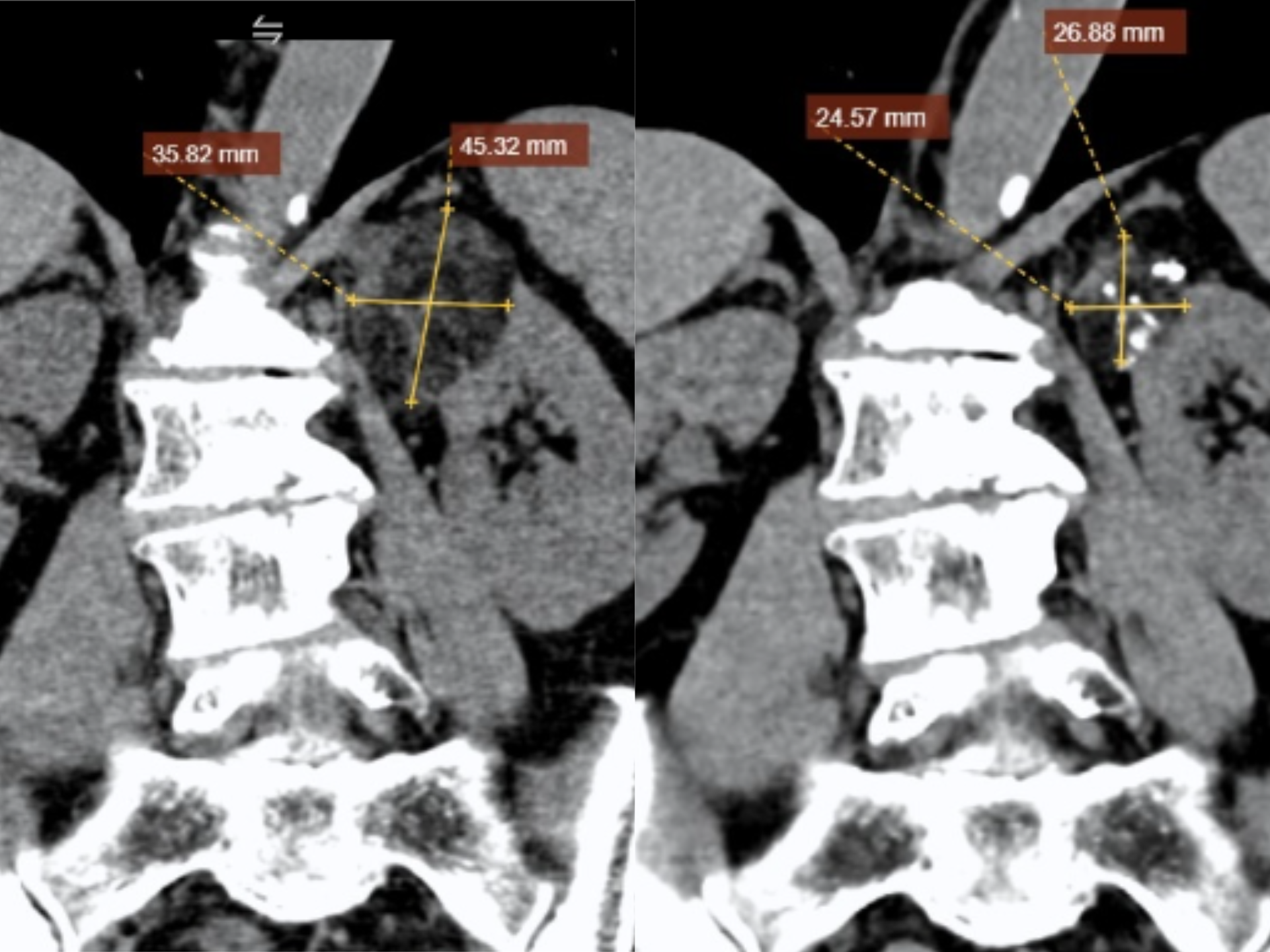 |
Ms. Hong's tumor size significantly decreased after 6 months of embolization. *Photo: Tam Anh General Hospital* |
Doctors advised treating Ms. Hong with tumor embolization guided by a digital subtraction angiography (DSA) system. After local anesthesia, the intervention team accessed the right femoral artery and guided a catheter through the vascular system to the left renal artery. Angiography images showed the tumor was primarily supplied by a branch of the left renal artery.
The team used a specialized microcatheter to selectively access the vessel branch feeding the tumor. Doctors then injected microspheres combined with biological glue to completely cut off the blood supply to the tumor, while preserving the vascular system supplying the healthy renal parenchyma.
 |
Associate Professor Hien inquired about Ms. Hong's health before her discharge. *Photo: Tam Anh General Hospital* |
After the intervention, Ms. Hong's health stabilized; she walked normally after 8 hours and was discharged after one day. Follow-up imaging after 6 months showed the tumor had shrunk by nearly half its size compared to before embolization.
Previously, patients with kidney angiomyolipomas often required partial nephrectomy, leading to loss of kidney function and reduced quality of life. Now, embolization assisted by the digital subtraction angiography (DSA) system helps doctors successfully treat large tumors without surgery, with fewer complications and a shorter recovery time.
Associate Professor Hien advised patients not to be complacent about symptoms such as persistent flank pain, hematuria, or a heavy sensation in the abdomen. Regular health check-ups can help detect kidney tumors at an early stage, allowing for appropriate treatment selection and limiting the risk of dangerous complications.
Hieu Nguyen
| Readers can ask questions about cancer here for doctors to answer |