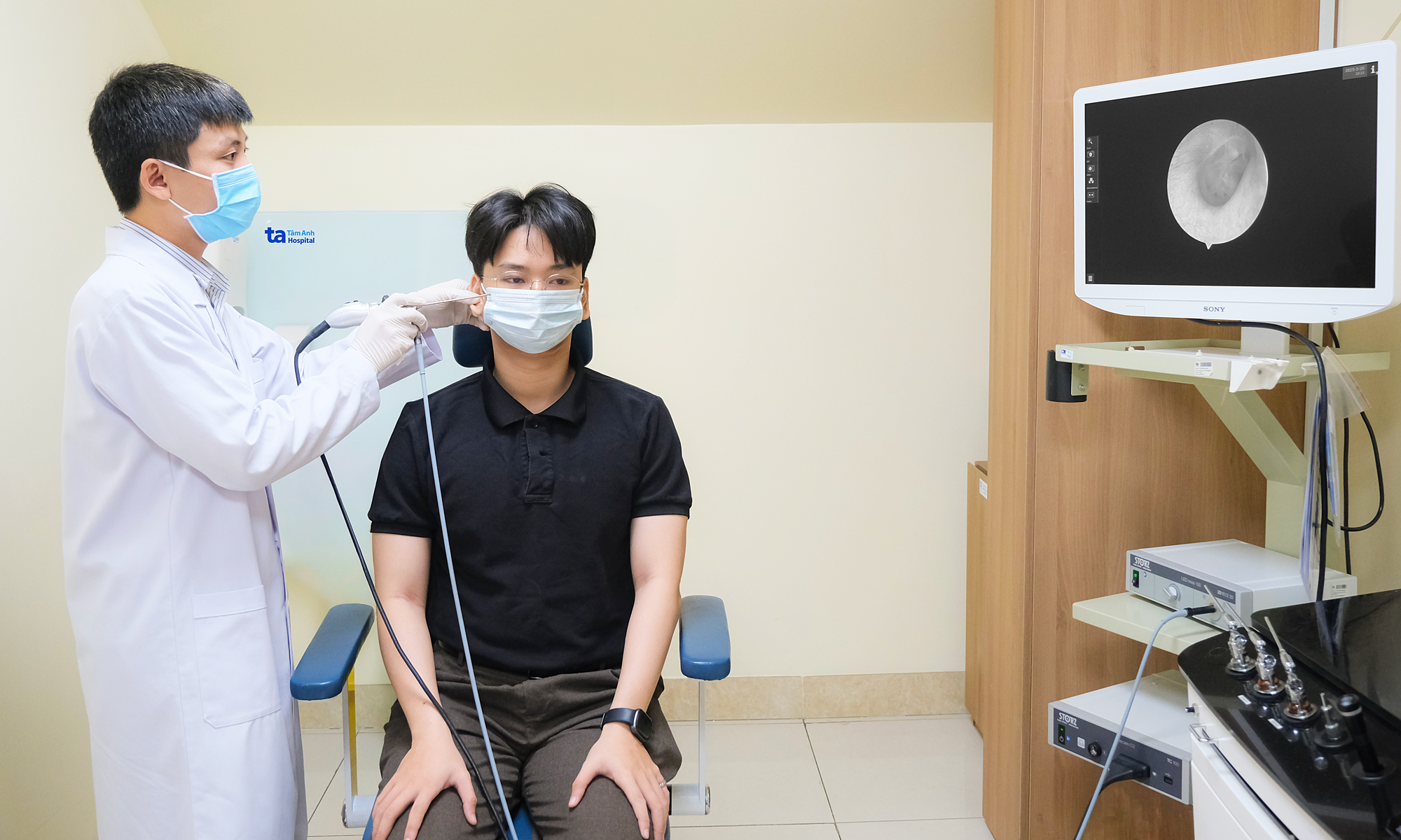
Ms. Phuong's MRI scan at Tam Anh Hanoi General Hospital revealed multiple lesions in her D12, L1, L2, L3, L4, and S1 vertebrae, indicating metastatic cancer. Specifically, the L1 vertebra was destroyed and collapsed, compressing her spinal cord, which caused her prolonged lower back pain.
A subsequent CT scan identified a 19x25 mm tumor in the lower left lung lobe and a nodule in the right liver. A biopsy of the spinal bone confirmed that Ms. Phuong had lung cancer that had metastasized to her bones, liver, and brain.
Master, Doctor Nguyen The Thu from the Oncology Department at Tam Anh Hanoi General Hospital, explained that Ms. Phuong's tumor was located in the periphery of the lower left lung lobe, close to the pleura and mediastinum. This peripheral location meant it caused few respiratory symptoms and was difficult to detect with standard chest X-rays.
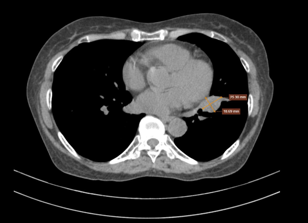 |
Ms. Phuong's tumor on a computed tomography scan. *Photo: Tam Anh General Hospital* |
Ms. Phuong had no history of smoking, coughing, or fever, though her father had suffered from lung cancer. Doctor Thu emphasized that not all lung cancer cases present with common symptoms like coughing or shortness of breath. Many early-stage tumors, particularly those in the lung's periphery, develop silently without affecting the airways. In such instances, symptoms like back pain, resulting from bone metastasis, can manifest before any respiratory issues.
The biopsy results showed Ms. Phuong had non-small cell lung cancer with an EGFR mutation. Doctor Thu noted that EGFR mutations are common among non-smokers, occurring more frequently in women than men. This mutation accounts for approximately 20% of adenocarcinomas in Western populations and 50-60% in Asian populations.
Ms. Phuong received systemic targeted therapy combined with medication to protect her bones and prevent bone destruction. The targeted therapy specifically inhibits the abnormal EGFR protein, thereby blocking signals that stimulate cancer cell proliferation and spread.
She responded well to the medication. After three months, the lesions in her lung and brain had significantly reduced in size. Following nearly two years and 22 treatment cycles, the lung tumor was no longer detectable on computed tomography scans, and bone regeneration was observed post-treatment.
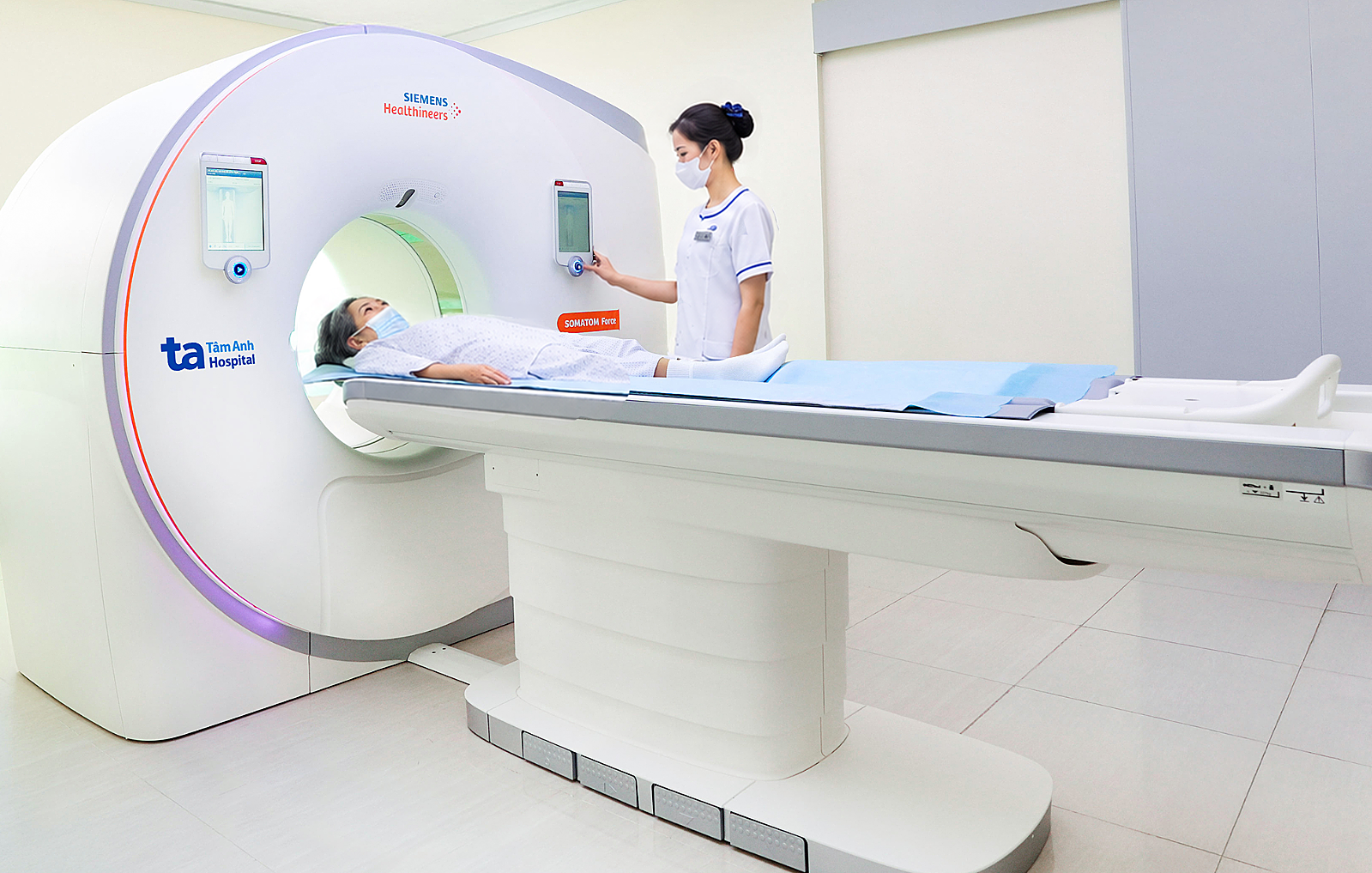 |
Ms. Phuong undergoing a low-dose CT scan using a Somatom Force VB30 machine for follow-up after nearly two years of treatment. *Photo: Tam Anh General Hospital* |
While smoking is the primary risk factor for lung cancer, other contributors include regular exposure to radioactive radon gas, toxic substances like chromium, nickel, cadmium, asbestos, and coal smoke.
Individuals who have undergone chest radiation, have a history of cancer, or have immediate family members (parents, children, siblings) who have had the disease are advised to undergo lung cancer screening as recommended by a doctor. Early detection through screening can lead to radical treatment of malignant lung nodules still localized within the lung tissue, often eliminating the need for adjuvant chemotherapy or radiation therapy.
Hieu Nguyen
| Readers can send questions about respiratory diseases here for doctors to answer |