

Mr. Son experienced abdominal pain, bloating, and fatigue for nearly two years. He was diagnosed with gastritis, but medication did not alleviate his symptoms. Recently, his pain intensified, accompanied by indigestion and constipation, prompting him to seek examination at Tam Anh General Hospital, TP HCM.
An abdominal ultrasound revealed a stomach wall thickening of approximately 8 mm in the pyloric antrum, along with an inflammatory mass infiltrating the fat. Master, Doctor Nguyen Thanh Bien, from the Center for Endoscopy and Endoscopic Digestive Surgery, identified a linear structure, suspected to be a foreign object, extending to liver segment III, near the head of the pancreas.
A CT scan confirmed a liver lesion measuring 4,5x3,8 cm, containing a foreign object nearly 4 cm long that had penetrated from the stomach wall into the liver parenchyma. Additionally, a smaller foreign object, 0,2 cm, was found in the sigmoid colon, though it had not yet perforated the colon wall.
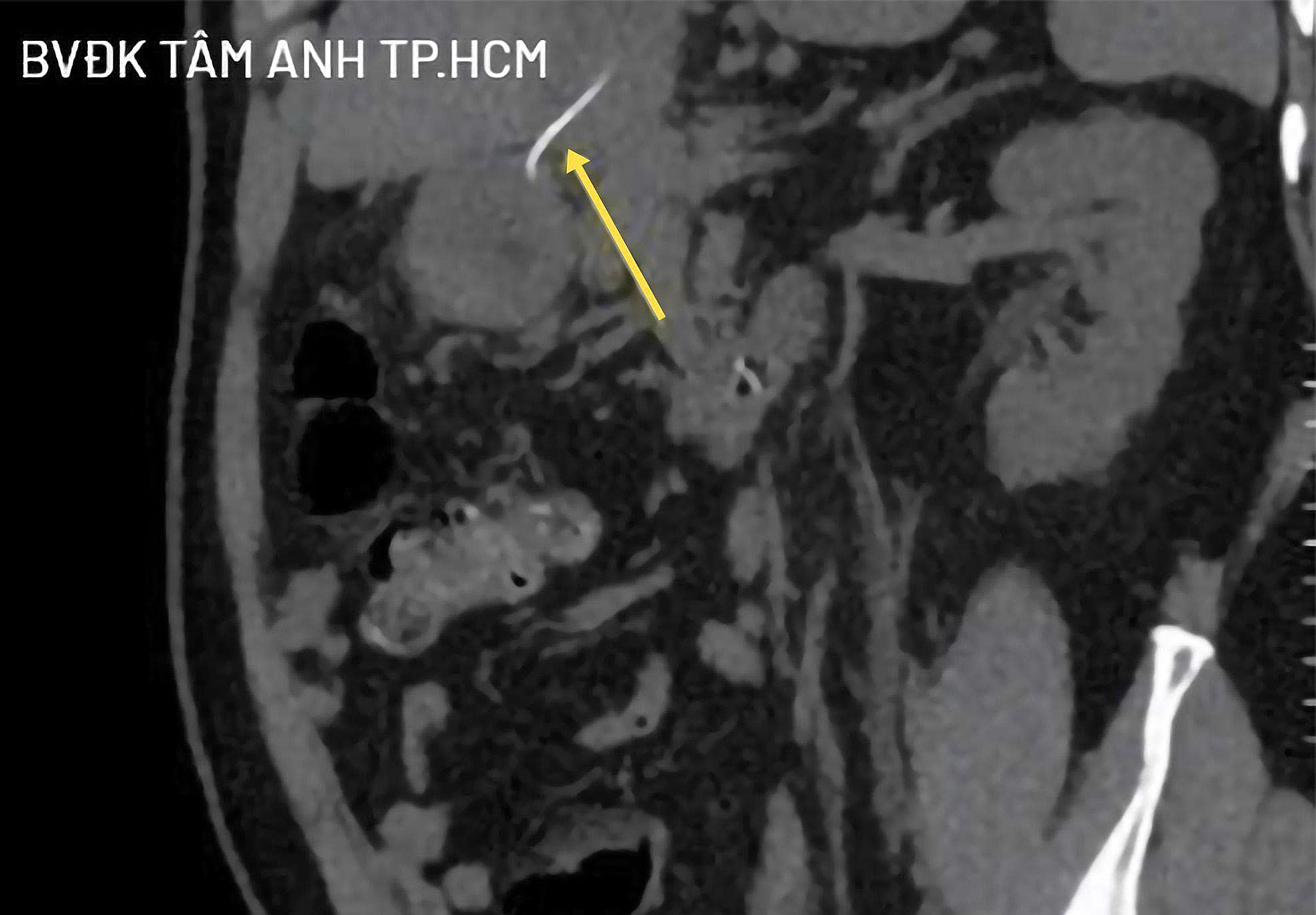 |
CT images show the foreign object penetrating the stomach wall into the liver parenchyma (yellow arrow). Photo: Tam Anh General Hospital |
"The foreign object had been in the body for a long time, mixed with necrotic inflammatory tissue, making detection difficult", Doctor Bien stated.
Mr. Son was scheduled for laparoscopic surgery to remove the foreign object. Due to his underlying conditions of hypertension and coronary artery disease, and his use of anticoagulant medication, the patient had to discontinue his medication five days prior to surgery and undergo a pre-operative physical assessment.
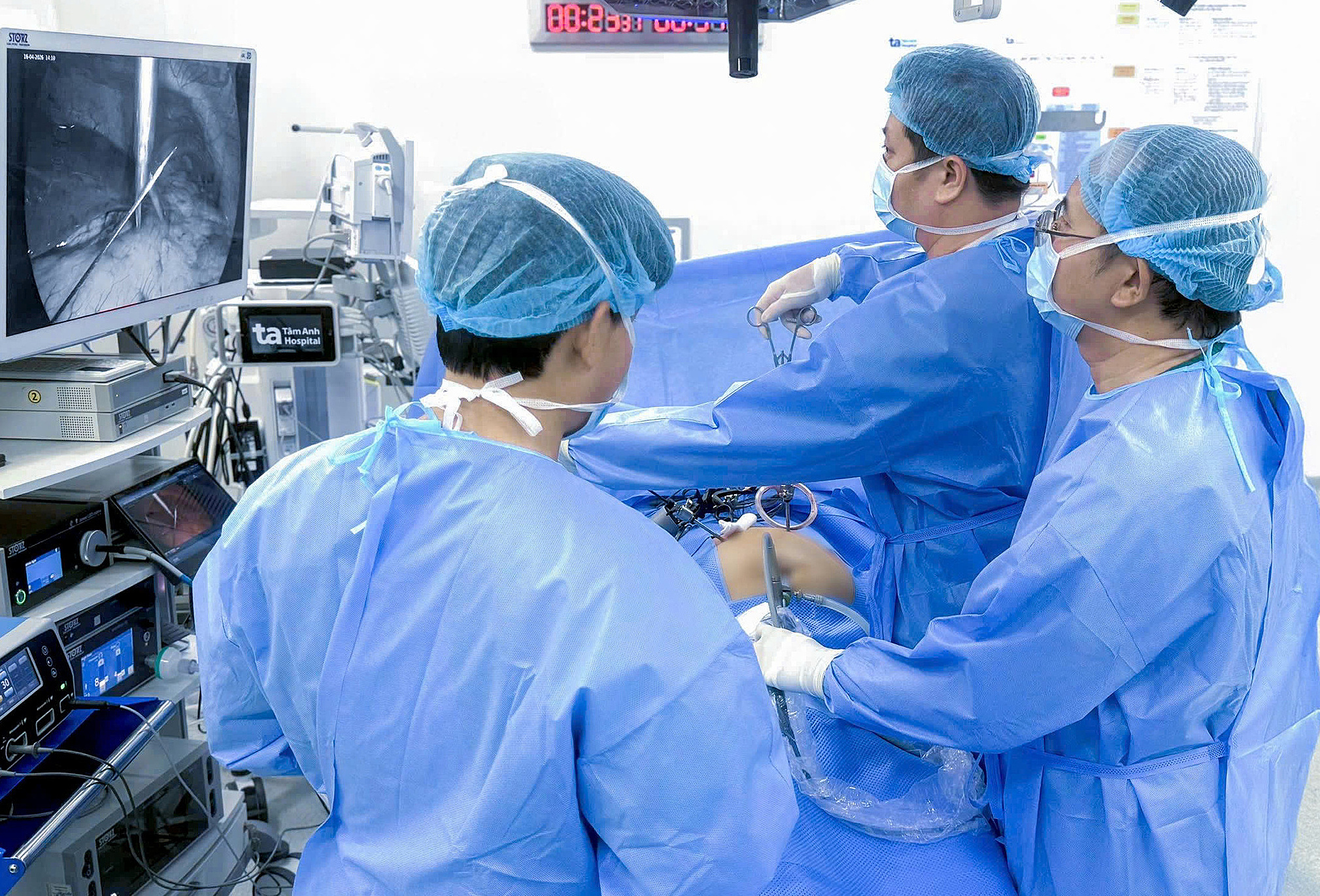 |
Doctor Bien (second from right) performs laparoscopic surgery to remove the foreign object for Mr. Son. Photo: Tam Anh General Hospital |
During the surgery, the medical team noted swelling and firm adhesion in the lesser curvature of the stomach, tightly bound to the left liver. Doctors discovered a fibrotic inflammatory mass containing a foreign object, suspected to be a nearly 4 cm long fish bone with two sharp ends, which had penetrated from the stomach into the liver parenchyma, with most of the object located within the liver. Doctors carefully removed the foreign object and re-examined the stomach.
Post-surgery, the patient experienced reduced abdominal pain, was able to consume liquids from day two, and was discharged after five days.
Swallowing foreign objects is a common household accident, frequently occurring in young children or elderly individuals. These objects vary widely, including bamboo toothpicks, toys, and bones from fish, chicken, or duck. While most foreign objects entering the digestive tract are naturally expelled through stool within a few days without complications, sharp or large foreign objects carry a high risk of perforating the digestive tract or causing intestinal obstruction, leading to numerous dangerous complications.
Once a foreign object has exited the digestive tract, gastroscopy and esophagoscopy may not detect it. A foreign object remaining in the abdominal cavity for an extended period can cause inflammation, leading to the formation of an abscess in the liver or adjacent organs.
Doctor Bien advises people, especially the elderly, to eat slowly and chew thoroughly when consuming foods with many small bones. He also recommends avoiding talking or using phones while eating, and refraining from holding toothpicks in the mouth. If a fish bone is suspected to have been swallowed, do not use folk remedies, as this can increase the risk of the foreign object penetrating tissues. If pain, a feeling of obstruction, or discomfort in the neck, chest, or epigastric region (above the navel) occurs, individuals should seek medical attention early for endoscopic removal of the foreign object; early intervention is safer and less invasive.
Quyen Phan
| Readers can submit questions about digestive diseases here for doctors to answer. |


