Ms. Thuy first discovered the hemangioma 10 years ago during surgery for a herniated disc. At that time, it was identified as an abnormal, benign tissue in the vertebral body that did not require intervention. Recently, she experienced severe back pain, numbness, and paralysis in both legs.
Dr. Tran Quang Hien, Head of Spinal Surgery at Tam Anh General Hospital Ho Chi Minh City, stated that the patient's hemangioma had grown significantly, measuring approximately 18x43,2 mm within the L4 vertebral body (which has a diameter of about 35-40 mm). It had almost completely destroyed this vertebra, severely compressing the dura mater and nerve roots.
Muscle strength examination revealed partial paralysis in both legs, with muscle strength in her left leg at only 2/5 and in her right leg at 3/5. The patient urgently needed surgery to prevent the risk of complete paralysis and sphincter dysfunction affecting bowel and bladder control.
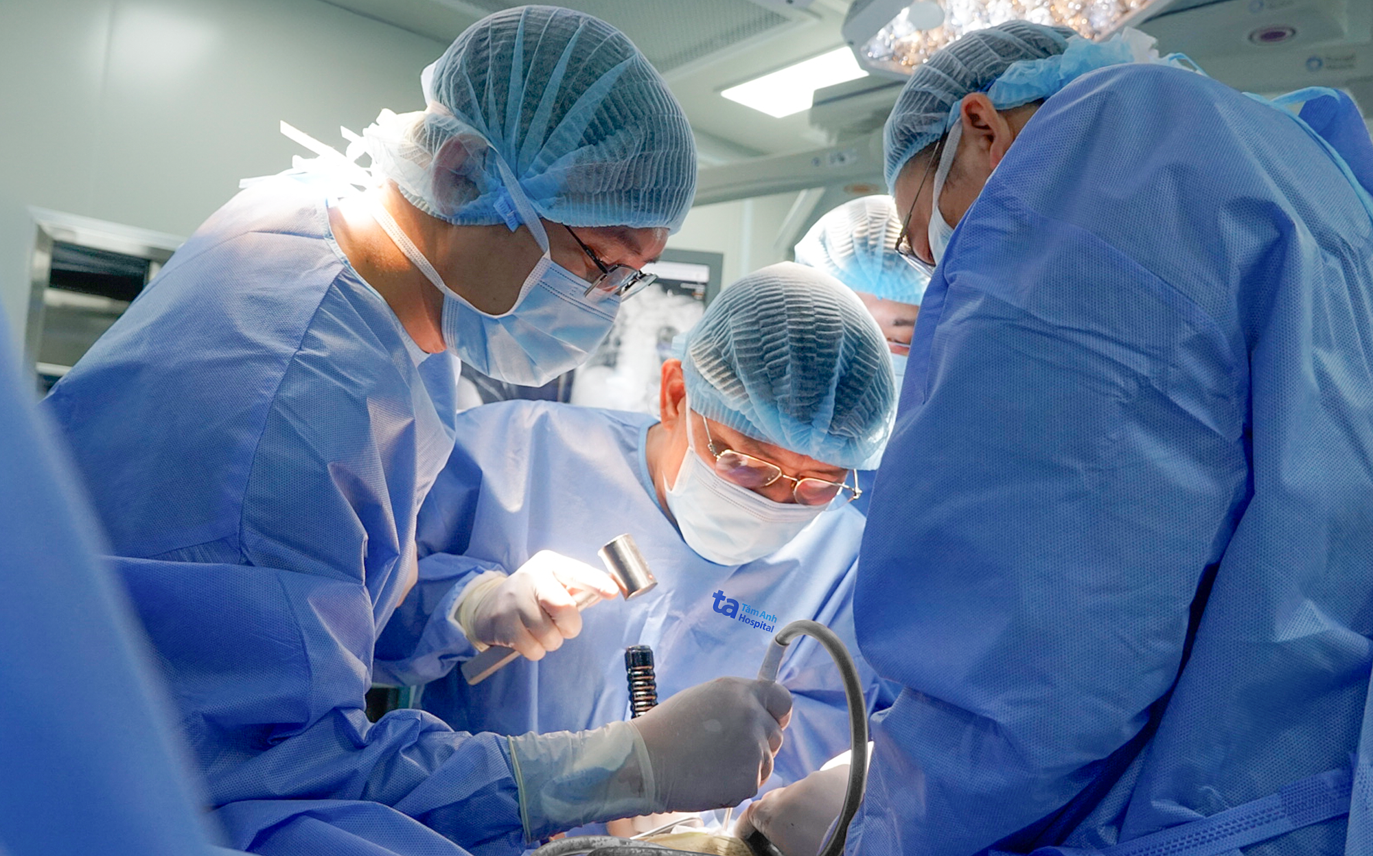 |
Dr. Hien (center) dissects the tumor for the patient. Photo: Tam Anh General Hospital |
Dr. Hien prescribed spinal fixation surgery and interbody fusion from the L2 lumbar vertebra to the first sacral vertebra S1. Due to the large size of the hemangioma, if not properly controlled, the patient faced a high risk of shock from significant blood loss and even death on the operating table.
The surgical team planned to control bleeding using bone wax (a sterile mixture of white beeswax and softening agents, used for mechanical hemostasis on damaged bone surfaces) and small absorbent gauze pads directly compressed onto the bleeding site. Ms. Thuy also suffered from diabetes and high blood pressure, increasing the risk of infection and further bleeding. Therefore, the surgery needed to be performed continuously to mitigate these potential risks.
To decompress the spine, doctors had to remove the L4 vertebra. However, this procedure could destabilize the spine and lead to severe consequences such as chronic pain, spinal deformity, and nerve damage. The patient's spine was stabilized with screws on one side, and the L4 vertebra was replaced with an expandable titanium cage to support the spine. Given her previous herniated disc surgery years ago, a significant amount of fibrous tissue had adhered tightly to the nerve roots. This made dissection and other maneuvers difficult, posing a high risk of nerve root damage leading to weakness and paralysis, demanding meticulous precision from the surgeons.
Ms. Thuy's blood loss was optimally managed, but she still required blood transfusions during surgery, post-operatively, and in the intensive care unit (ICU). On the 5th day after surgery, the patient was able to walk steadily with a frame and no longer needed a wheelchair.
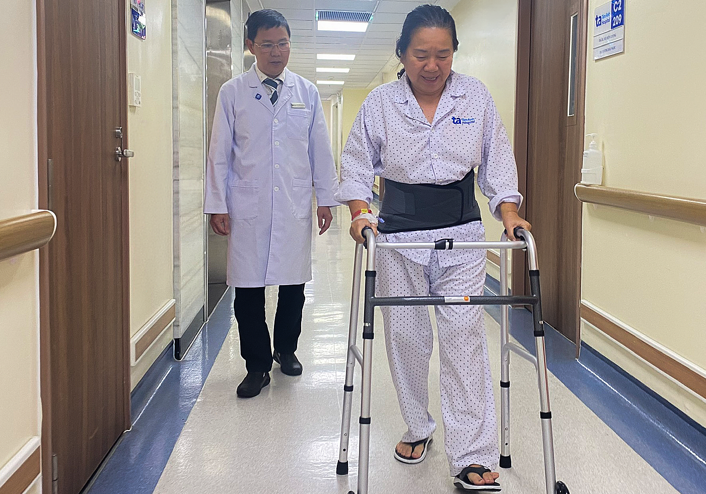 |
Dr. Hien guides the patient in walking after surgery. Photo: Tam Anh General Hospital |
Dr. Hien noted that the hemangioma had deeply adhered to the spinal cord, and despite the successful surgery, Ms. Thuy would find it challenging to recover completely to her pre-illness state. However, by adhering to the treatment plan and rehabilitation exercises, the patient could restore mobility and experience minimal impact on her daily activities.
Vertebral hemangiomas are not rare; they are typically benign and asymptomatic. To limit their growth, doctors may employ various methods: embolize the blood vessels feeding the tumor, inject biological cement into the vertebral body, or perform radiation therapy.
Doctors advise individuals diagnosed with a hemangioma to undergo regular health check-ups to monitor the condition, assess the tumor's growth rate and size, and intervene promptly to prevent complications.
Phi Hong