An echocardiogram of Ms. Nghiep at Tam Anh General Hospital Hanoi revealed severe pulmonary hypertension, with an estimated systolic pulmonary artery pressure of 65 mmHg. The patient's right ventricle was significantly dilated, right ventricular systolic function was reduced, and tricuspid regurgitation was present.
Doctor Le Ngoc Anh, from the Cardiology Department, diagnosed Ms. Nghiep with pulmonary hypertension due to a connective tissue disease. This diagnosis was supported by positive anti-ANA, Centromere A, Centromere B, Sp 100, and PML test results. Connective tissue disease is an autoimmune condition where the immune system mistakenly attacks the body's own tissues, leading to inflammation and damage in multiple organs. Autoantibodies can damage the pulmonary vascular bed, resulting in fibrosis, vascular obstruction, endothelial dysfunction, and impaired vascular regulation. These changes increase pulmonary artery pressure, overloading the right ventricle and causing right heart failure, which manifests as patient fatigue and shortness of breath.
Ms. Nghiep also had several underlying conditions: hypertension, type 2 diabetes, moderate chronic coronary artery atherosclerosis, heart failure with preserved ejection fraction, and chronic kidney disease, all of which increased her risk of complications.
 |
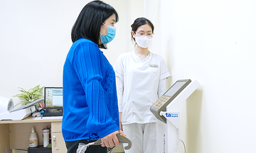
Doctor Ngoc Anh examining Ms. Nghiep. *Photo: Tam Anh General Hospital* |
Doctors prescribed a combination treatment: pulmonary artery pressure-lowering medication, connective tissue disease medication, and diuretics. One week later, her shortness of breath decreased, heart rate and blood pressure stabilized, coughing stopped, lung fluid cleared, and she was discharged.
She had a follow-up appointment three months later, where her health showed significant improvement. Her heart rate and blood pressure were stable, exertion-related fatigue symptoms reduced, systolic pulmonary artery pressure decreased to 35 mmHg, and heart size and function returned to normal.
According to Doctor Ngoc Anh, patients with pulmonary hypertension often receive a late diagnosis, after severe symptoms or target organ damage have already appeared. Many cases are not correctly classified into the appropriate pulmonary hypertension group, leading to unsuitable treatment. Determining the cause, diagnosis, and treatment requires multidisciplinary coordination among cardiology, respiratory, rheumatology, immunology-allergy, intensive care, and imaging diagnostics specialists.
Pulmonary hypertension is categorized into main groups: pulmonary arterial hypertension, pulmonary hypertension related to left heart disease, lung disease or hypoxia, and pulmonary hypertension due to pulmonary artery obstruction. Treatment plans vary depending on the underlying cause.
Doctors recommend screening and diagnosing pulmonary hypertension in individuals with connective tissue diseases such as scleroderma, systemic lupus erythematosus, Sjogren's syndrome, those carrying the BMPR2 gene mutation, or first-degree relatives of patients with heritable pulmonary arterial hypertension. Patients undergoing pre-liver transplant evaluation, or with portal hypertension, HIV infection, unexplained shortness of breath, left heart disease, or chronic lung disease should also undergo examination.
Ly Nguyen
| Readers can submit questions about cardiology here for doctors to answer |