Mr. Cuong suddenly experienced severe headaches, slow reactions, and difficulty walking after a flight. A computed tomography (CT) scan of the brain at Tam Anh General Hospital Hanoi revealed a large amount of air had leaked into the skull due to a fistula at the skull base.
The patient previously suffered a traumatic brain injury, skull fracture, and bilateral brain contusions. Master of Science, Doctor Le Huu Tung, from the Department of Neurosurgery – Spine, stated that the patient underwent prolonged intensive care and received a ventriculoperitoneal shunt due to hydrocephalus. Post-surgery, the patient recovered well, with motor skills, communication, and daily activities returning almost to normal for over one year.
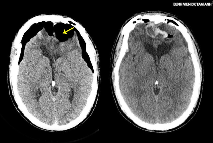 |
Mr. Cuong's intracranial air accumulation before and after surgery. *Photo: Tam Anh General Hospital*
Doctor Tung noted that many patients recover from injuries and live normal lives for extended periods. However, areas of the skull base with incomplete healing from bone defects, dural scars, or damage remain "weak points."
Under normal conditions, these sites maintain a seal between the intracranial cavity and the paranasal sinuses, so patients remain asymptomatic. However, minor impacts like pressure changes during air travel, head and facial trauma, forceful sneezing, nose blowing, prolonged coughing, or a slight bump can compromise these weak points. This creates a pathway between the intracranial cavity and the external environment, allowing air from the paranasal sinuses to enter the skull.
In many cases, the fistula acts as a "one-way valve," allowing air to enter but not escape. Excessive air accumulation leads to tension pneumocephalus, compressing brain tissue and increasing intracranial pressure, which is life-threatening, according to Doctor Tung.
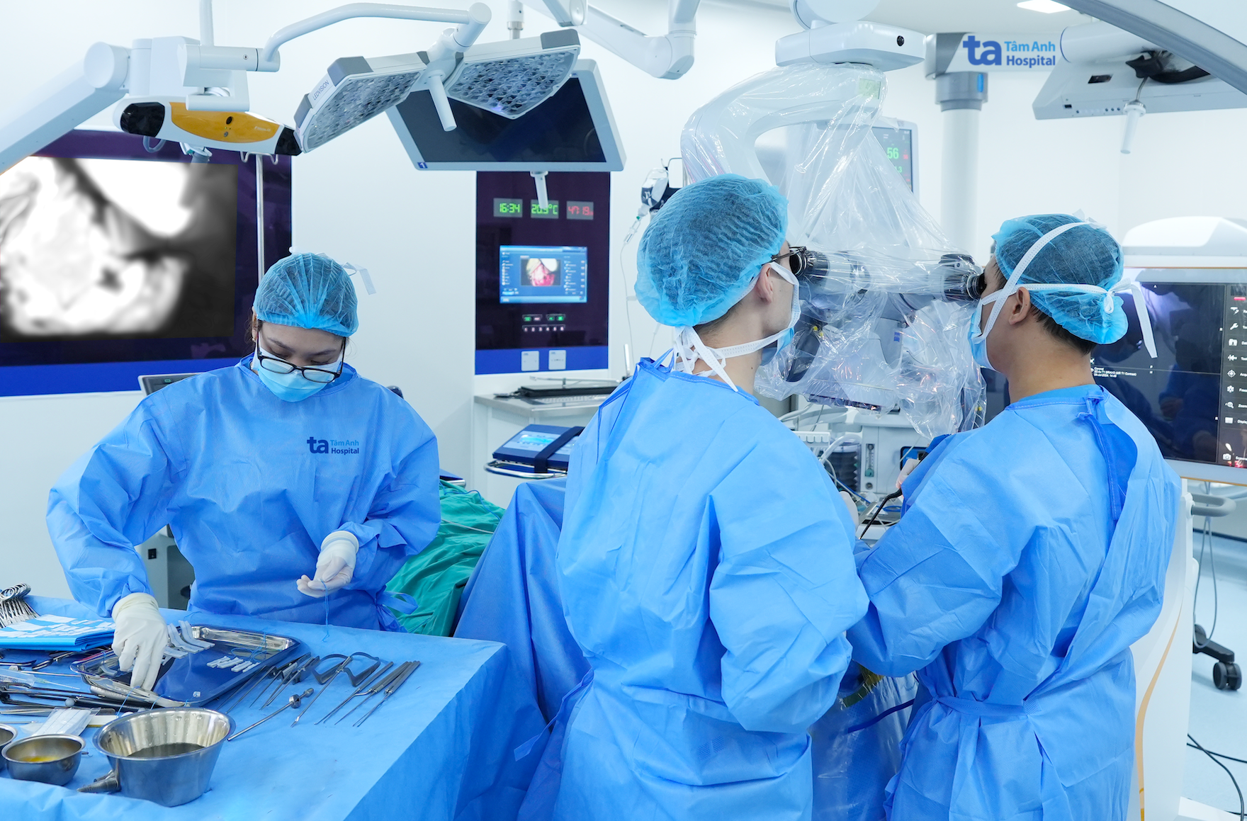 |
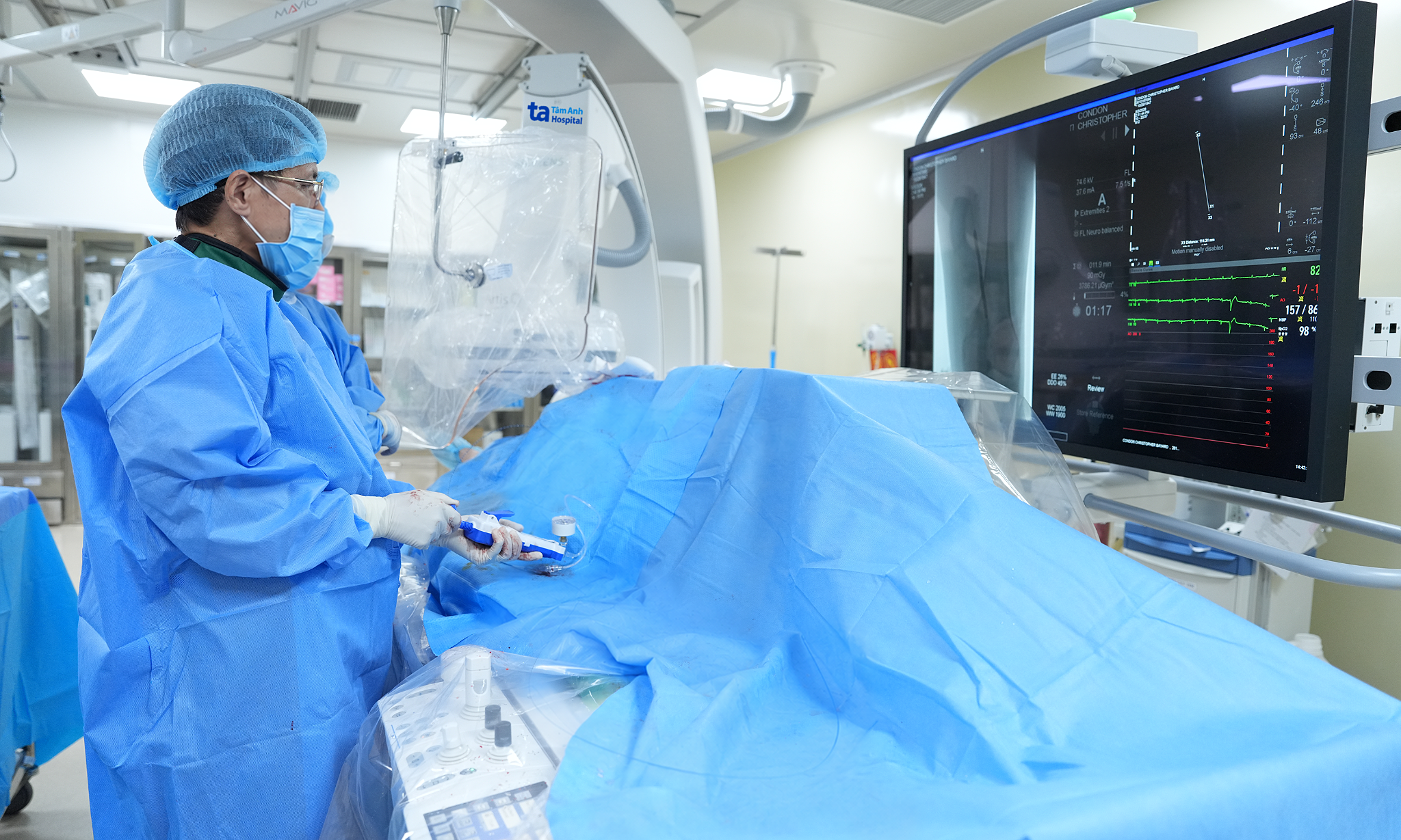
Doctor Tung (center) performs surgery on Mr. Cuong. *Photo: Hospital provided*
Doctors decided on craniotomy for decompression and skull base reconstruction for Mr. Cuong.
The surgical team used a microscope to identify two fistulas at the bilateral frontal skull base, communicating with the frontal sinuses. Surgeons utilized autologous fat and fascia lata to seal the dural tears, combining biological glue and dural patch material to reconstruct the skull base in multiple layers, preventing communication between the cranial cavity and the air-filled sinuses.
Following surgery, the air in the skull was released, the fistulas were sealed, and the patient recovered well, with improved consciousness, able to walk again, and discharged after one week of treatment.
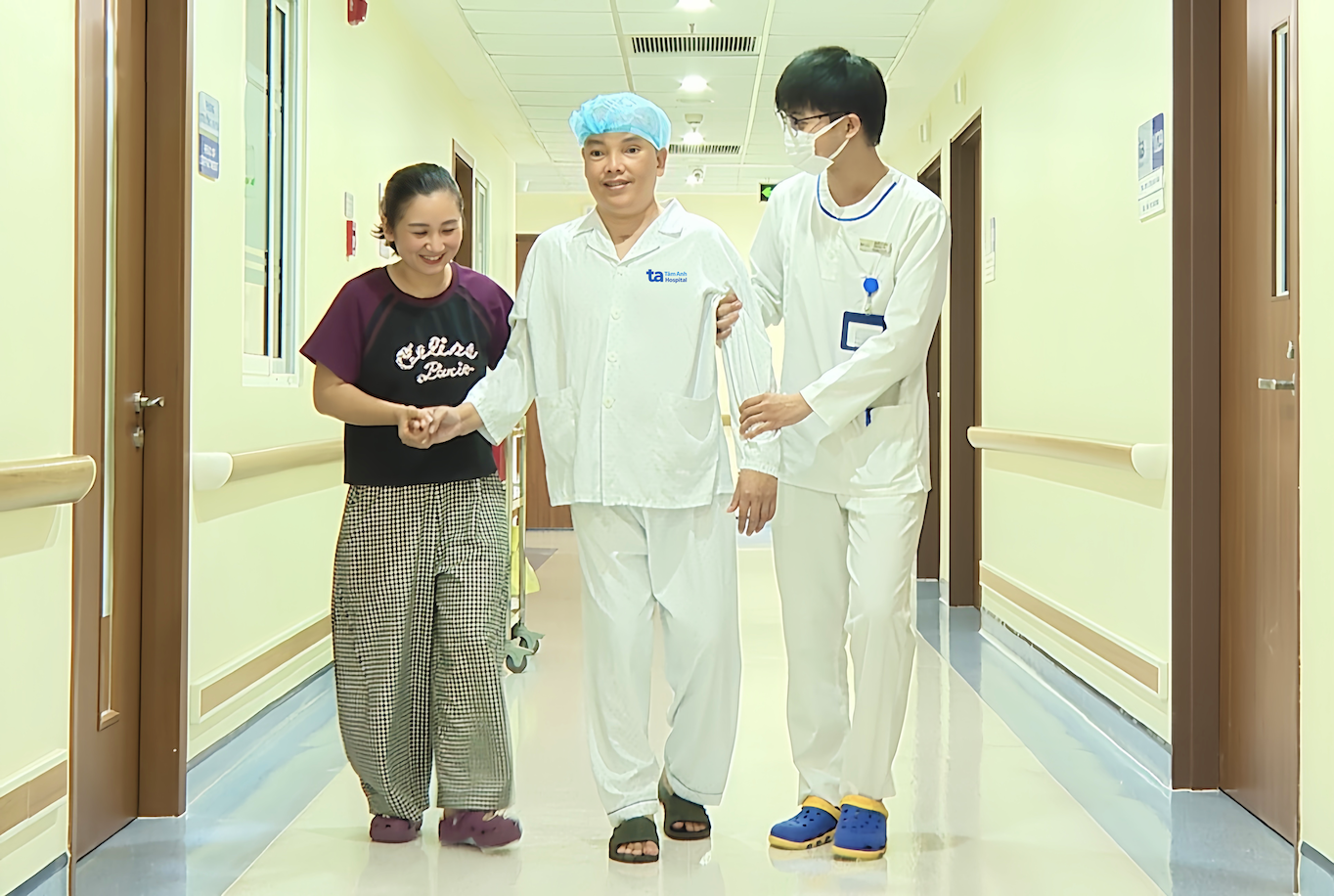 |
Mr. Cuong regained the ability to walk after surgery. *Photo: Tam Anh General Hospital*
Doctor Tung added that not all patients with a history of traumatic brain injury are prohibited from flying. However, individuals with a history of skull base fracture, cerebrospinal fluid leak, skull base surgery, skull base bone defects after trauma, or those who have had a ventriculoperitoneal shunt need careful risk assessment by a specialist before long-haul flights.
If symptoms such as progressive headaches after a flight, nausea, decreased alertness, limb weakness, or hearing sloshing sounds inside the head occur, patients should seek early diagnosis and treatment at a hospital with a neurosurgery department. Doctors recommend that individuals with a history of traumatic brain injury or skull base surgery, even if recovered, still need regular follow-ups to detect abnormalities and protect long-term brain function.
Thanh Long
| Readers can ask questions about neurological diseases here for doctors to answer. |