During a general health check-up at Benh vien Da khoa Tam Anh TP HCM, Ba Thu, 59, who had returned from the US to visit relatives, received a surprising diagnosis. An abdominal ultrasound revealed a large ovarian tumor measuring 13 x 13 x 13,5 cm. Additionally, the examination identified multiple uterine fibroids, polyps, and an abnormally thick uterine lining.
Given the tumor's size and Ba Thu's postmenopausal status, BS.CKI Ung Quoc Thuong from the Trung tam San Phu khoa, ordered biochemical tests for ovarian tumors. Although the results indicated a low malignancy risk of approximately 21% for postmenopausal women, a subsequent pelvic MRI showed images suspicious for a mature teratoma in the left ovary. The doctor recommended surgery to remove the tumor and conduct a comprehensive evaluation.
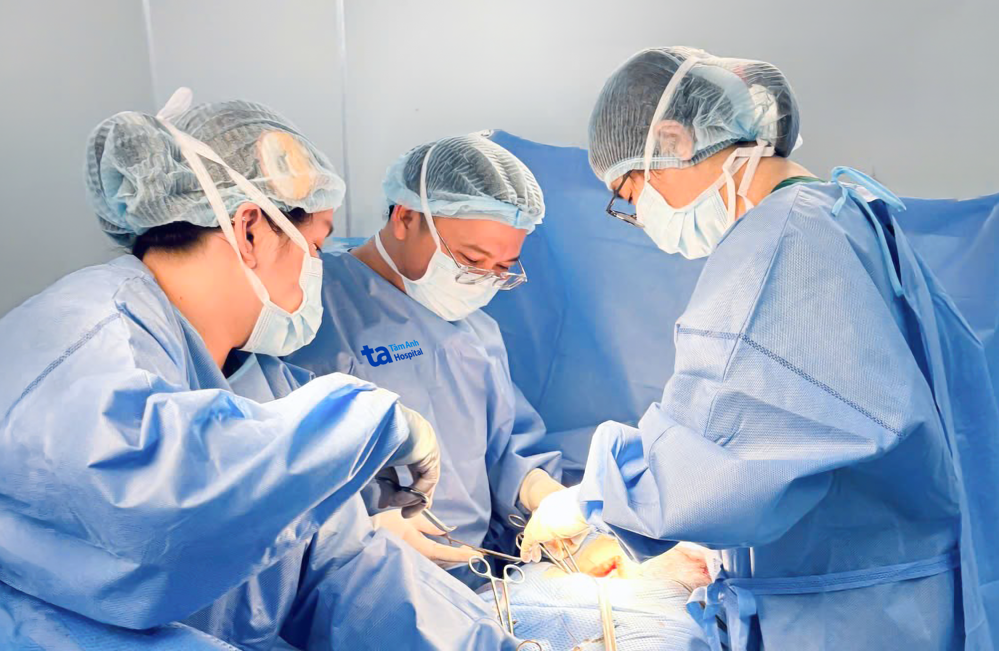 |
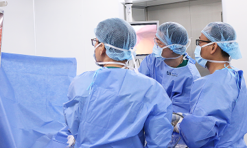
Doctor Thuong (center) and the Trung tam San Phu khoa team performed a total hysterectomy and oophorectomy for Ba Thu. *Photo: Benh vien Da khoa Tam Anh*
The surgical team successfully removed the entire uterus, both adnexa, and the ovarian tumor intact in a procedure lasting nearly three hours. Upon incising the tumor, doctors observed adipose tissue, hair, and fluid—components typically found in a teratoma. However, they also discovered multiple suspicious papillary masses, indicating a high likelihood of malignancy. Consequently, the team proceeded with radical surgery, which included an omentectomy and removal of the paracolic gutter peritoneum for further post-operative assessment.
Pathology analysis of tissue samples from the tumor, processed into thin slices and stained with H&E for microscopic examination, confirmed keratinizing squamous cell carcinoma arising from a mature teratoma, classified as stage T1a. The tumor was localized to the left ovary with an intact capsule, and no malignant cells were detected in the peritoneal fluid, omentum, or peritoneum.
Doctor Thuong explained that only about 0,17-2% of mature cystic ovarian teratomas develop into malignancy. Of these, 80% transform into squamous cell carcinoma, as seen in Ba Thu's case. He noted that such occurrences are rare.
"The initial tumor may have been a mature teratoma, but over time, an area of tissue within it underwent abnormal changes, leading to the formation of cancer cells," Doctor Thuong elaborated. This transformation process often progresses silently, with a higher risk in older, postmenopausal women.
Ba Thu's disease was detected at stage I, offering a significantly better prognosis compared to patients diagnosed at stages II, III, or IV. Following radical surgery, she will continue to receive monitoring from gynecologists and oncologists.
Ovarian cancer is a common gynecological cancer, ranking after breast and cervical cancer, and is associated with a high mortality rate and poor prognosis. While the exact cause remains unclear, risk factors may include genetics, reproductive history, and environmental influences. The disease often progresses silently, making early detection challenging.
Doctor Thuong advises women to be vigilant for symptoms such as gradual abdominal enlargement, bloating, abdominal pain, indigestion, constipation, back pain, unexplained weight loss, fatigue, frequent or involuntary urination, abnormal uterine bleeding, and abdominal distension. He recommends regular gynecological check-ups, ultrasounds, and other diagnostic tests or imaging when indicated.
Tue Diem
| Readers can send their questions about obstetrics and gynecology here for doctors to answer. |