Vuong first discovered a pituitary tumor 6 years ago. The tumor continuously recurred, leading to three previous surgeries at a local hospital. Recently, his vision deteriorated, prompting him to seek examination at Tam Anh General Hospital Hanoi.
Master of Science, Doctor Le Huu Tung, from the Neurosurgery - Spine Department, noted that Vuong exhibited signs of bilateral optic disc necrosis, severe visual field loss, and color blindness. His right eye failed to perceive light signals. Optical coherence tomography (OCT) revealed optic nerve atrophy. An MRI scan further confirmed a giant pituitary adenoma, measuring 5x5x4 cm, alongside multiple lesions and hematomas.
According to Doctor Tung, pituitary tumors with a diameter greater than 4 cm are classified as giant. Vuong's tumor had invaded deep brain regions, reaching the third ventricle and obstructing the flow of cerebrospinal fluid. The tumor displaced brain tissue and compressed the optic chiasm, resulting in a complete loss of signals in some visual pathways.
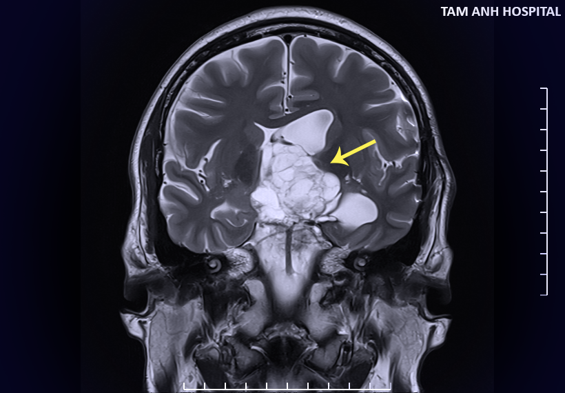 |
Large tumor in Vuong's pituitary gland. Photo: *Tam Anh General Hospital*. |
Following a multidisciplinary consultation, doctors recommended surgery to remove the tumor. The surgical team utilized a neuro-navigation system to create a 3D anatomical reconstruction of the tumor, pituitary gland, and entire brain. Intraoperative neuromonitoring was also employed to detect any early risks of nerve damage, ensuring maximum functional preservation for the patient during the procedure.
Doctor Tung explained that the tumor's large size and the patient's history of three previous surgeries had caused fibrous adhesions in the brain structures, making dissection challenging. The surgical team opted for a lateral ventricular approach, a more complex technique than the conventional transsphenoidal approach for pituitary tumor removal. This method, however, provided better access for complete resection of giant pituitary tumors that had invaded the ventricles.
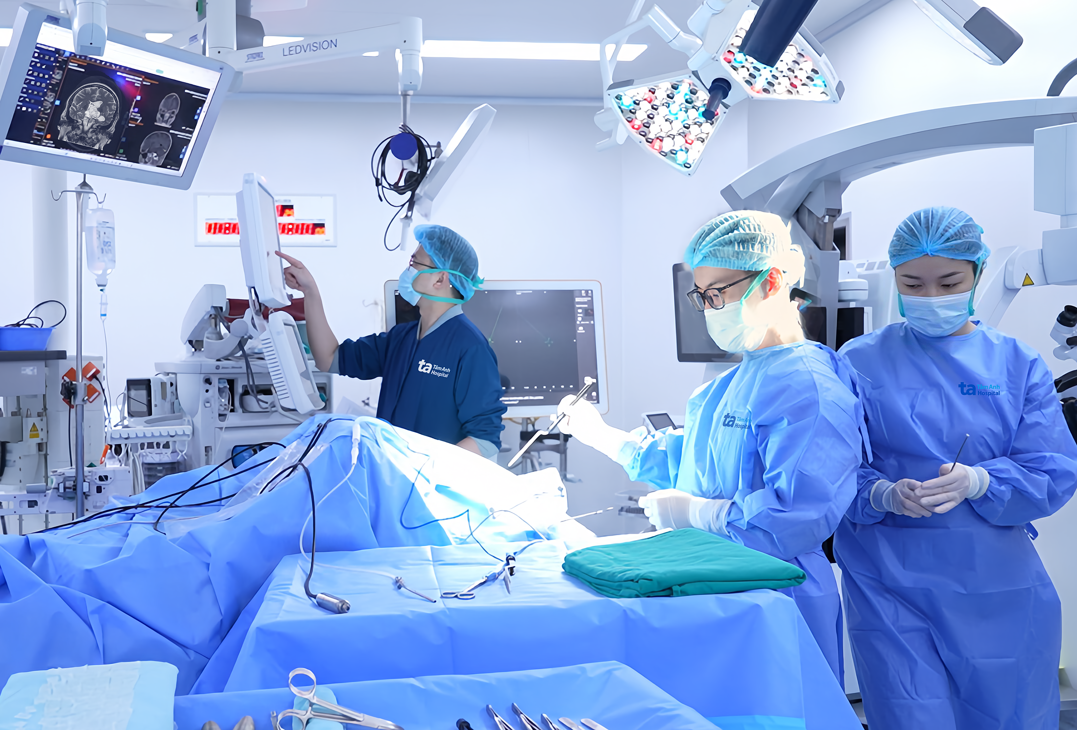 |
Doctor Tung (center) performs surgery on Vuong. Photo: *Hospital provided*. |
Under an AI-powered microsurgery microscope and 3D fluorescence imaging system, doctors clearly observed the tumor's boundaries relative to the healthy pituitary tissue that needed preservation. This allowed for precise, partial tumor resection, minimizing invasion.
Post-surgery, Vuong was alert, his vision gradually restored, and the incision was dry. He was discharged after 7 days. At a follow-up appointment one month later, his visual field had fully improved, and his color blindness had resolved.
 |
Vuong recovers vision and mobility after surgery. Photo: *Tam Anh General Hospital*. |
The pituitary gland is an endocrine gland located at the base of the skull. It produces and releases essential hormones that regulate growth, metabolism, reproductive function, and the activity of other endocrine glands.
Normally, the pituitary gland is small, about the size of a pea. Pituitary tumors develop when pituitary cells proliferate abnormally. According to Doctor Tung, most pituitary tumors are benign and non-cancerous, meaning they do not metastasize. However, giant tumors can compress surrounding structures, leading to symptoms such as: headaches, vision loss, infertility, and cognitive decline.
Common symptoms of pituitary tumors include: headaches, vision loss, lethargy, extreme fatigue, frequent urination, and excessive thirst. Patients experiencing these symptoms should visit a hospital with a neurosurgery department for thorough examination and treatment.
Thanh Long
| Readers can send questions about neurological diseases here for doctors to answer. |