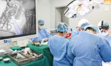
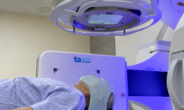
Tam Anh General Hospital Hanoi activated a "code red," establishing a priority pathway to prepare for Mr. Long's emergency surgery. Initial cranial CT scans revealed a left frontal lobe abscess, ventricular dilation, and altered cerebrospinal fluid signals.
Master of Science, Doctor Bui Khuong Duy, deputy head of the Neurosurgery and Spine Department, diagnosed Mr. Long with intraventricular ruptured brain abscess (IVROBA). This is one of brain abscess's most dangerous complications. The cerebral ventricles are a system of cavities deep within the brain that contain and circulate cerebrospinal fluid. When an abscess ruptures, pus and bacteria spread with the cerebrospinal fluid to many areas of the central nervous system, causing ventriculitis, meningitis, and widespread infection.
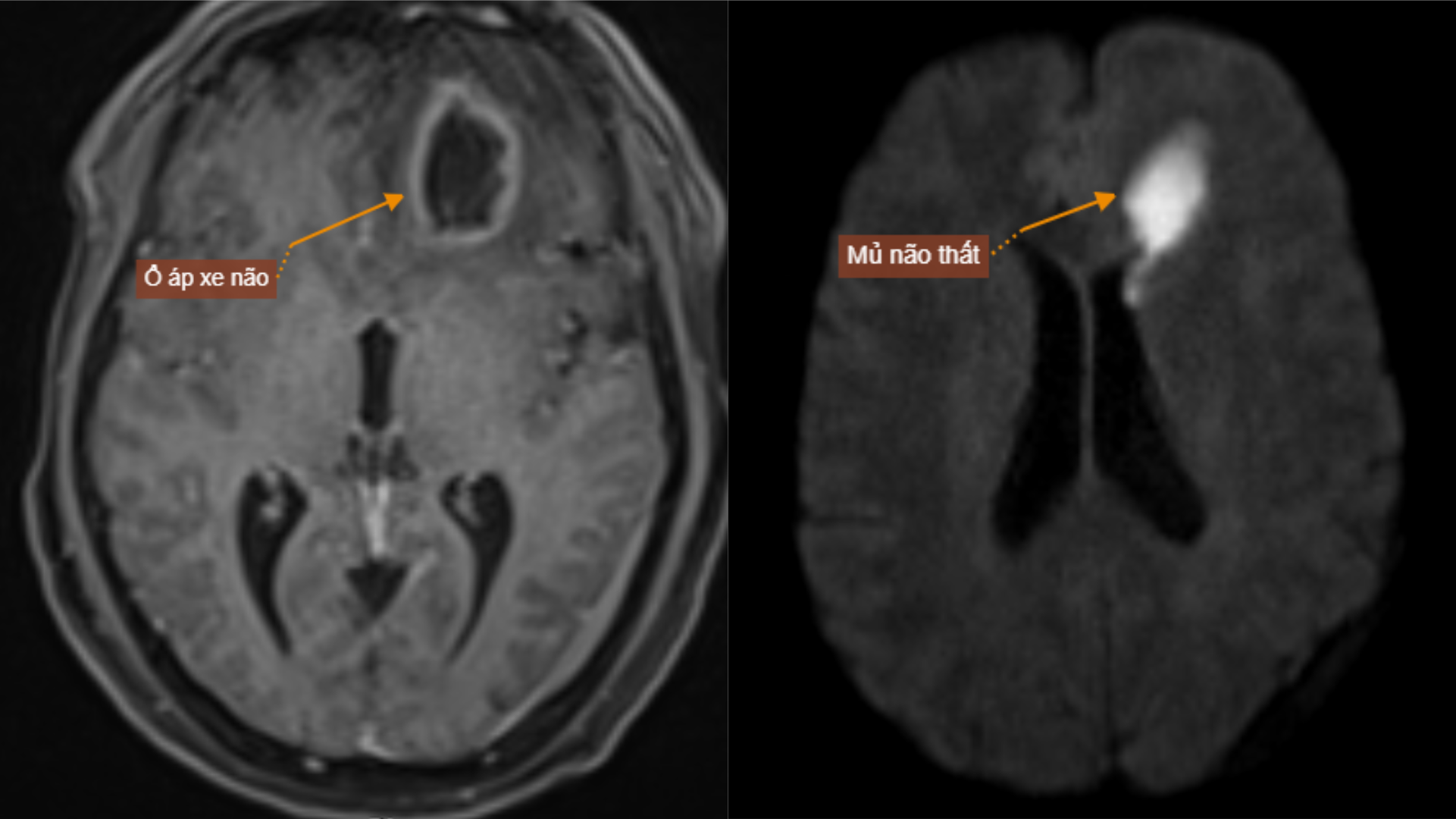 |
Images show Mr. Long's ruptured brain abscess, forming pus in the cerebral ventricles. *Photo: Tam Anh General Hospital* |
One month prior, Mr. Long suffered a traumatic brain injury that caused a skull base fracture, creating a fistula from the maxillary sinus into the skull. According to Doctor Duy, this provided an entry point for bacteria, leading to the formation of an abscess in the left frontal lobe and its subsequent rupture into the cerebral ventricles.
The patient underwent minimally invasive surgery (intervention through small incisions) for external ventricular drainage combined with abscess aspiration. After locating the lesion, doctors accessed the abscess in the left frontal lobe, aspirating over 30 ml of turbid yellow pus, and simultaneously placed a catheter into the ventricle to drain cerebrospinal fluid. Following the first surgery, the patient received intensive care, including blood sugar and blood pressure control, and antibiotic treatment to manage the infection.
Two weeks later, with his condition improved, Mr. Long underwent a second surgery. Doctors performed a left frontotemporal craniotomy, exposed the skull base, and discovered an anterior skull base fracture with a dural tear, forming a communicating tunnel between the maxillary sinus region and the subdural space. Surgeons repaired the dura mater, re-establishing the barrier between the subdural space and the skull base, thereby eliminating the entry pathway for bacteria from the sinus region, then closed the incision.
Three days after the surgery, Mr. Long was conscious and his awareness improved. Four weeks later, the surgical wound healed well; he was speaking, eating, and walking normally, with no complications. Follow-up cranial MRI scans showed no ventricular dilation and no evidence of the abscess.
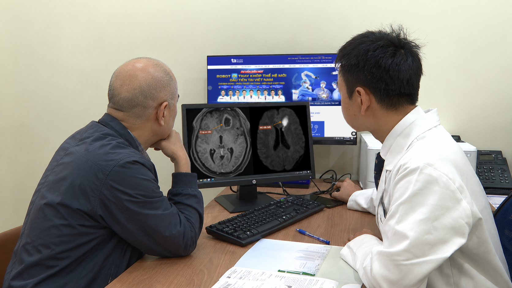 |
Doctor Khuong Duy advises Mr. Long on his post-surgery abscess condition. *Photo: Provided by the hospital* |
Doctor Khuong Duy stated that ruptured brain abscess complications have a high mortality rate, approximately 84%. Surgery is the primary treatment method for ruptured brain abscesses. Early detection and timely intervention before an abscess ruptures significantly help limit complications.
Following a traumatic brain injury, patients require monitoring and adherence to follow-up appointments. If they experience progressively worsening headaches, fever, vomiting, altered consciousness, seizures, or other unusual neurological symptoms, patients should seek medical attention for examination to rule out the risk of intracranial infection, brain abscess, and other dangerous neurological complications.
Hieu Nguyen
*Patient's name has been changed
| Readers can ask questions about neurological diseases here for doctors to answer. |