Meng had suffered from kidney stones for over 10 years, experiencing frequent renal colic, recurrent urinary tract infections, and requiring a JJ stent when stones obstructed her ureter. She sought examination at Tam Anh General Hospital, TP HCM. A CT scan revealed multiple upper renal calyx stones, the largest measuring 10-12 mm, along with an appendiceal mucocele measuring 58x25 mm in the right iliac fossa, which was leaking fluid.
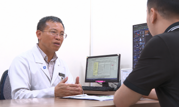
Dr. Ly Minh Hoang, from the Department of Urology, Nephrology, and Andrology, explained that Meng's kidney stones repeatedly recurred and were difficult to treat due to a congenital ureteral stricture, which impeded urine flow from the kidney to the bladder. When stones reached this narrowed area, the ureteral lumen became more prone to obstruction, causing hydronephrosis and fostering recurrent urinary tract infections. Additionally, the stricture made it challenging for standard endoscopic instruments to access the renal calyces and pelvis system, especially when stones were deeply embedded.
Dr. Vo Ngoc Bich, from the Department of Hepatobiliary-Pancreatic Surgery, Center for Endoscopy and Gastrointestinal Endoscopic Surgery, stated that an appendiceal mucocele can be benign or associated with various forms of cancer. As the tumor grows, the appendix wall can thin, risking leakage or rupture. If mucus and tumor cells spill into the abdominal cavity, the patient may develop pseudomyxoma peritonei, a complex condition requiring specialized treatment.
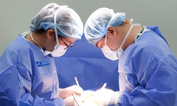
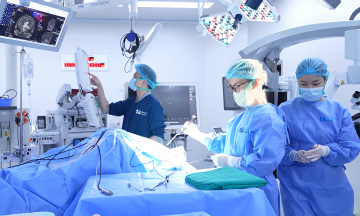
Meng's tumor required early intervention, but the surgical procedure had to prevent further damage or rupture of the appendiceal wall, which would release mucus into the abdominal cavity. After consultation, the surgical team decided to perform two operations under a single anesthetic: completely removing the kidney stones and excising the appendiceal mucocele.
In the first surgery, Dr. Hoang removed the old JJ stent and inserted a flexible endoscope into the urinary tract to perform lithotripsy. The instrument's flexible tip allowed it to navigate the narrowed ureteral segment and reach deep into the renal calyces and pelvis system, where the stone was lodged. The stone was fragmented by laser and then completely removed using a flexible 3 mm diameter sheath, preventing stone fragments from abrading the body during extraction.
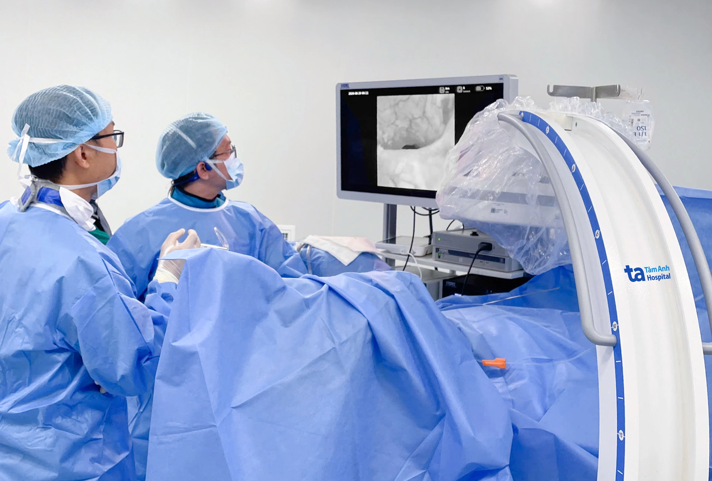 |
Dr. Hoang (right) uses a flexible endoscope combined with a laser for Meng's kidney stone lithotripsy. Photo: Tam Anh General Hospital |
Subsequently, the surgical team inserted endoscopic instruments into the abdominal cavity to address the appendiceal mucocele. During direct observation, doctors noted the tumor measured approximately 60x30 mm, larger than indicated by the CT scan. A rupture point, about 5x5 mm, was present at the tumor's distal end, actively secreting mucus but enveloped by the greater omentum.
Dr. Bich dissected the tumor, secured the appendiceal stump with three specialized clips, then placed the specimen into a bag and removed it through the navel incision, minimizing mucus exposure to the abdominal cavity. Meng recovered well and was discharged after 48 hours. Her follow-up appointment in early september showed her urinary system was stone-free and urine flow was good.
Appendiceal mucoceles often progress silently, causing no obvious symptoms, and may be discovered incidentally during a health check-up or a CT scan for another condition. Individuals experiencing unusual pain in the lower right abdomen or prolonged digestive disorders should seek medical evaluation. Early management of an appendiceal mucocele is crucial to limit the risk of leakage or rupture, which can lead to dangerous complications. Dr. Hoang advises that individuals with a history of recurrent kidney stones, renal colic, or multiple urinary tract infections should also undergo specialist examination.
Dinh Lam
*Patient's name has been changed
| Readers can ask questions about kidney disease here for doctors to answer |