Joint cartilage covers bone ends, creating a smooth surface for bones to glide over each other during movement. It also distributes forces applied to the joint. Cartilage consists of water, proteoglycans, and a tightly organized network of collagen.
Doctor Hoang Quyet Tien from the Medical Information Center at Tam Anh General Hospital Ho Chi Minh City states that collagen type II accounts for approximately 90-95% of the total collagen in adult joint cartilage. This is a key difference between collagen type I and type II. Collagen type I is common in tissues such as skin, tendons, ligaments, and bones, while type II is characteristic of cartilage. Each type is organized in a way that suits the properties of the tissue it forms.
 |
Joint cartilage structure is largely formed by collagen type II. Photo: Eco Pharmaceutical |
Joint cartilage structure is largely formed by collagen type II. Photo: Eco Pharmaceutical
According to doctor Tien, type II collagen fibers form a network surrounding the cartilage matrix, binding with proteoglycans. This helps organize and hold matrix components in a stable structure. While collagen provides strength to the network, proteoglycans can retain water and help cartilage resist compressive forces. The synergy between these two components gives cartilage both firmness and elasticity, allowing it to adapt to continuous forces on the joint.
Every day, joint cartilage endures force from walking, standing, sitting, climbing stairs, or other movements. To withstand these impacts, the collagen network in the cartilage must be strong enough to maintain tissue structure. When the links within the collagen network are preserved, cartilage retains its shape, strength, and load-bearing capacity.
In healthy cartilage, chondrocytes continuously synthesize and maintain matrix components. When osteoarthritis occurs, the breakdown of the matrix increases, including the degradation of collagen type II.
Certain enzymes, such as MMP-1 and MMP-13, can cleave collagen type II. When the collagen network is damaged, the matrix structure gradually changes, affecting the mechanical properties of the cartilage.
This change causes cartilage to progressively lose its firmness and ability to adapt to impacts. Its capacity to protect and distribute forces for the bone ends is also compromised, which can lead to pain, joint stiffness, and restricted movement.
 |
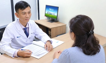
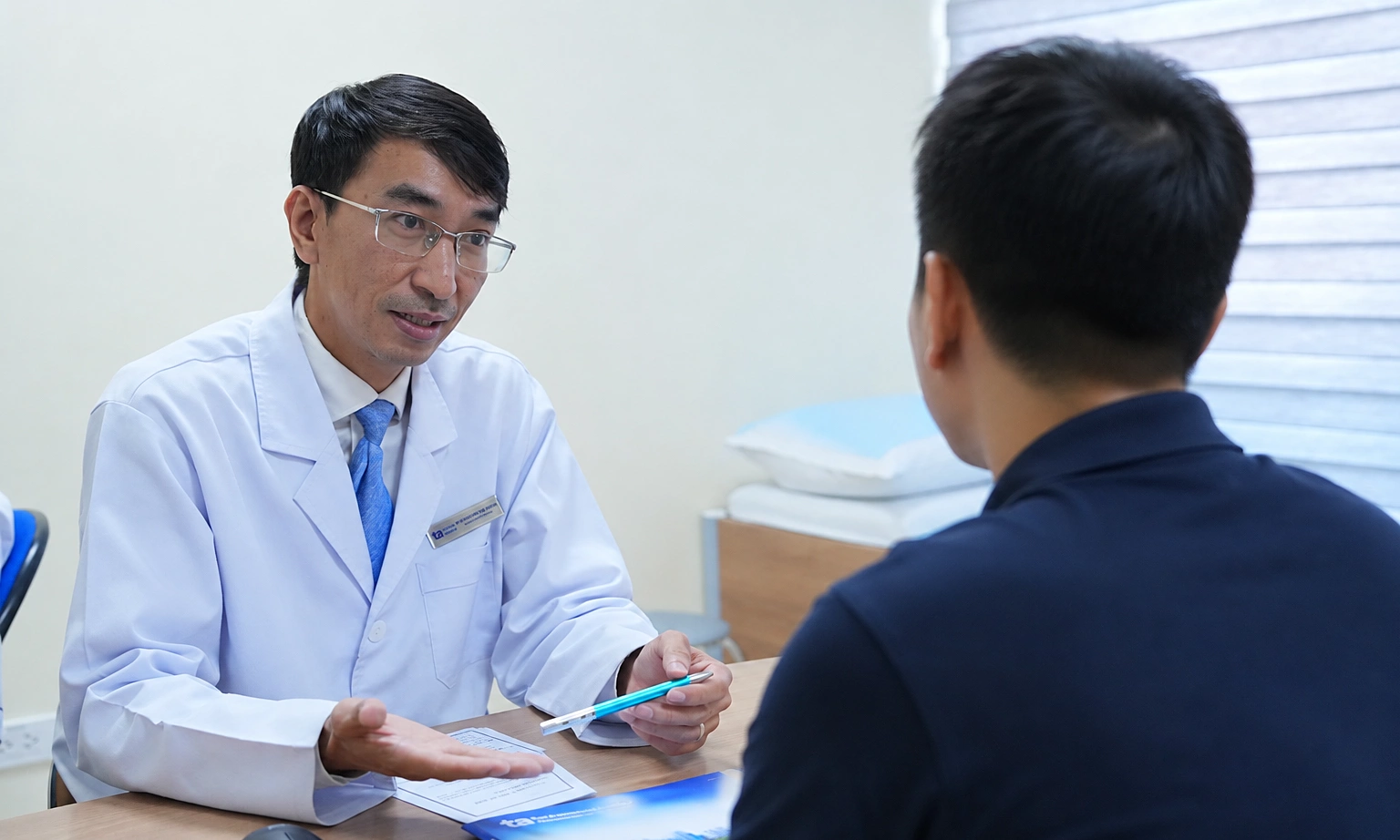
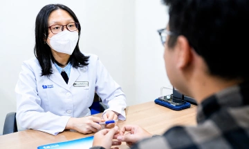
A doctor at Tam Anh General Hospital consults a patient. Photo: Hospital provided |
A doctor at Tam Anh General Hospital consults a patient. Photo: Hospital provided
Doctor Tien emphasizes that collagen type II, especially undenatured collagen type II (UC 2), is a crucial component in building joint cartilage. UC 2 is highly effective in reducing pain and joint stiffness, helping patients with knee osteoarthritis walk and move more easily. Supplementing with UC 2 supports immune modulation, protects joint cartilage and synovial membrane, and regenerates cartilage.
Doctor Tien advises everyone to care for their joints early, especially those who are regularly active, perform heavy work, or begin to experience pain and joint stiffness. Alongside maintaining a healthy weight and appropriate exercise, supplementing with natural nutrients such as the combination of undenatured collagen type II and collagen peptide, eggshell membrane, chondroitin sulfate, and turmeric extract also supports joint cartilage regeneration, pain reduction, and maintenance of motor function.
Dinh Dieu
| Readers can send questions about musculoskeletal diseases here for a doctor's answer. |