Epilepsy is a neurological disorder characterized by abnormal brain activity. This leads to the simultaneous excitation of a group of cortical nerve cells, causing sudden, uncontrolled electrical discharges.
Symptoms include tonic-clonic seizures or focal seizures affecting one body area, such as an arm or leg, often accompanied by foaming at the mouth. Patients may also experience sudden absence seizures, where they stop activity for 3-30 seconds, stare blankly, and then return to normal. The frequency of these symptoms varies among individuals.
Doctor Nguyen Duc Anh, Head of Neurosurgery - Spine Department at Tam Anh General Hospital Hanoi, identifies 5 common brain lesions associated with epilepsy.
Focal cortical dysplasia is a developmental abnormality of the cerebral cortex originating from the fetal stage. It occurs when nerve cells migrate and arrange incorrectly. This affected cortical region generates abnormal electrical signals in the brain, which can spread and trigger recurrent epileptic seizures. The condition often manifests in childhood and frequently proves resistant to medication.
Hypothalamic hamartoma is a congenital, benign tumor located in the brain's hypothalamic region. It forms from abnormally developed nerve tissue that can spontaneously generate electrical impulses. This can lead to characteristic epileptic seizures, most notably uncontrolled laughter. The condition typically begins in early childhood and may also be associated with behavioral disorders or precocious puberty.
Hippocampal sclerosis is a common cause of temporal lobe epilepsy. This condition involves the atrophy and scarring of the hippocampus, leading to nerve cell death. This instability in the brain's electrical activity then triggers epileptic seizures.
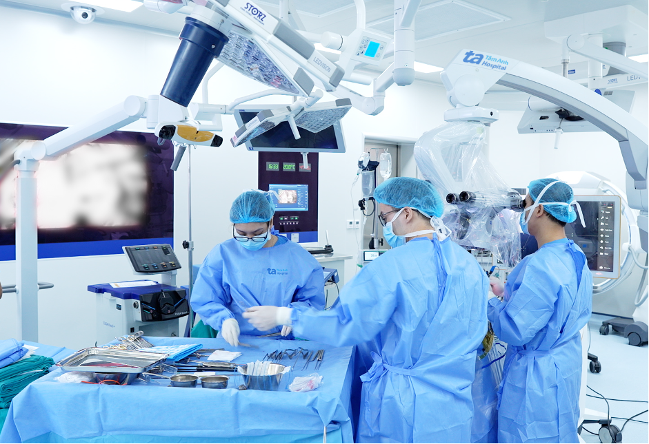 |
Surgeons remove a brain tumor to treat epilepsy. *Photo: Tam Anh General Hospital*
Brain vascular malformations describe conditions where cerebral blood vessels develop abnormally. This can result in tangled vascular masses or other aberrant vascular structures. These structural changes irritate surrounding brain tissue, which can trigger epileptic seizures.
Dysembryoplastic neuroepithelial tumor (DNET) is a rare, benign brain tumor. It forms from the abnormal development of nerve and glial cells during embryonic development. Though DNETs are slow-growing and rarely malignant, they are strongly linked to chronic epilepsy, particularly seizures that are difficult to manage with medication. DNET tumors are typically found in the cerebral cortex, most often in the temporal lobe.
According to Doctor Duc Anh, many epilepsy patients face challenges in diagnosis, as conventional methods often fail to identify the underlying cause. This complicates treatment, often necessitating long-term medication to control seizures and increasing the risk of drug resistance.
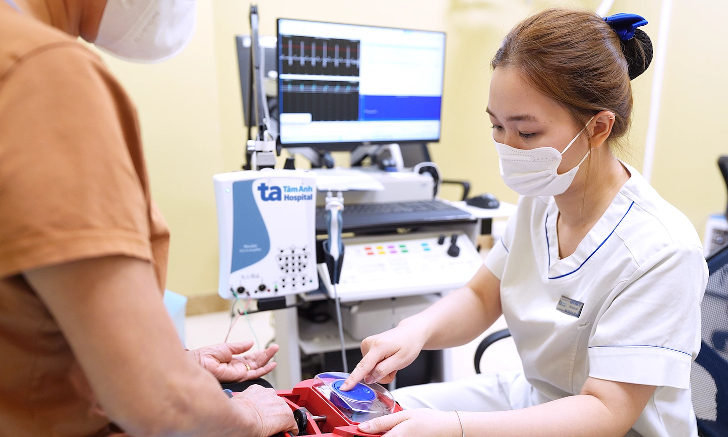
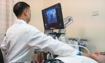
In certain cases, doctors utilize advanced diagnostic imaging tools for epilepsy, including 3.0 Tesla magnetic resonance imaging (MRI) and functional MRI. These are combined with specialized exploratory techniques like video electroencephalography (EEG), stereoelectroencephalography (SEEG), and electrocorticography (ECoG) to pinpoint brain lesions responsible for seizures.
Epilepsy caused by structural brain lesions can be definitively treated if the precise seizure-generating brain region is identified and the lesion is surgically accessible. Doctors perform comprehensive evaluations, including brain functional mapping, before surgically removing the lesion. This intervention can help patients achieve seizure freedom or significantly reduce seizure frequency.
Thanh Long
| Readers can submit questions about neurological diseases here for expert answers. |