Upon receiving information from Mr. Minh's family, Tam Anh General Hospital Ho Chi Minh City immediately activated its Code Stroke protocol. This dedicated emergency pathway for stroke patients significantly reduces the time required for assessment and treatment.
A cerebral computed tomography (CT) angiography revealed a 6x5 mm aneurysm at the A2-A3 segment of his anterior cerebral artery. The aneurysm's irregular border and the presence of blood under the meninges in both frontal regions confirmed subarachnoid hemorrhage.
Dr. Mai Hoang Vu, Head of the Neuro-Spine Surgery Department at the Neuroscience Center, stated that this was a critical situation. The lesion's location, surrounded by vital blood vessels supplying the brain, posed significant risk. The ruptured aneurysm, marked by a small tear, carried a high risk of re-rupture, which could lead to massive brain bleeding, deep coma, or death.
Following a multidisciplinary consultation, surgeons decided to perform an aneurysm clipping procedure on Mr. Minh. They utilized an AI-integrated microsurgical microscope system with 3D fluorescence imaging, allowing real-time blood flow monitoring during the operation.
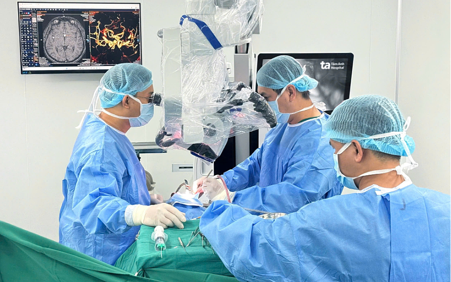 |
Doctors performing aneurysm clipping on Mr. Minh using the K.Zeiss Kinevo 900 AI microsurgical system. Photo: Tam Anh General Hospital |
According to Dr. Vu, the patient's aneurysm was deep within the interhemispheric fissure, close to numerous small brain-supplying vessels. The aneurysm wall was thin and had already ruptured, increasing the risk of severe bleeding. Before surgery, a neuro-navigation system generated a 3D brain image, guiding the team to identify a safe access route and track instrument positions in real-time.
After opening the dura mater, doctors used the microsurgical microscope to carefully trace the anterior cerebral artery branches, approaching the aneurysm. They performed slow, gentle maneuvers to avoid touching small brain-supplying vessels and the aneurysm wall. The aneurysm was meticulously separated from surrounding connective tissues, preserving the small arterial branches.
Once the aneurysm neck was clearly identified, the team placed a specialized titanium clip across it. Fluorescent dye was injected intravenously, and blood flow was observed directly through the microsurgical microscope. Images confirmed good blood circulation through the parent artery and its small branches, while the aneurysm no longer received blood supply, indicating complete treatment, Dr. Vu explained.
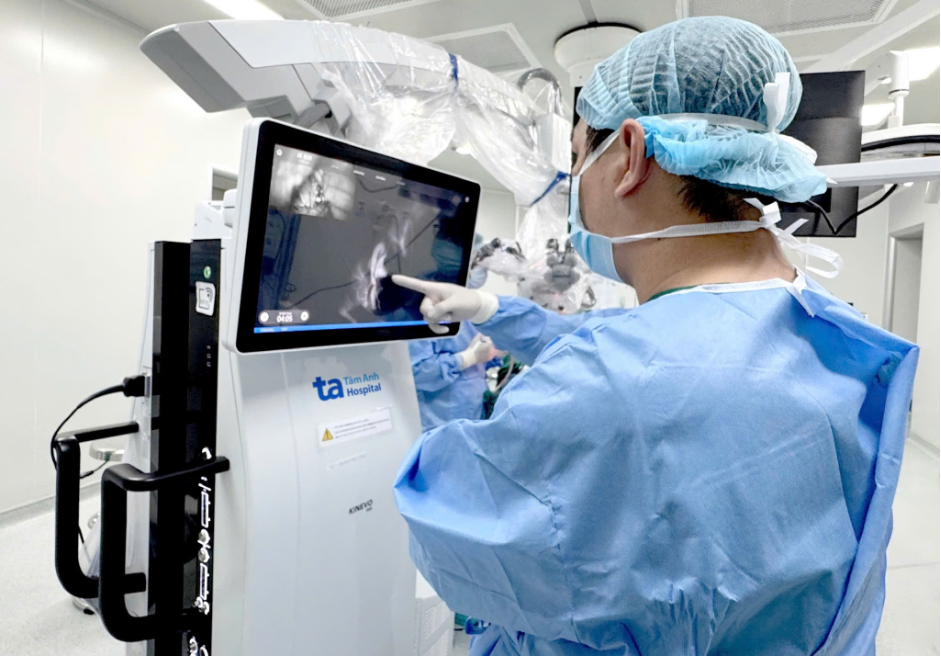 |
Dr. Vu checking blood circulation using the microsurgical microscope's fluorescence imaging technique after aneurysm clipping. Photo: Tam Anh General Hospital |
Three days post-surgery, Mr. Minh was alert. He could sit up and began walking with assistance from physical therapists to restore his strength and motor function.
Dr. Vu explained that a brain aneurysm occurs when a blood vessel wall weakens and bulges like a "balloon." Most aneurysms show no clear symptoms until they rupture. Some cases present warning signs such as persistent dull headaches, blurred vision, drooping eyelids, or pain around the eye socket. High-risk groups include individuals with hypertension, smokers, those who consume alcohol, people with a family history of cerebrovascular disease, or those previously diagnosed with an aneurysm.
Doctors advise seeking medical attention immediately for any sudden, severe, unusual headache accompanied by nausea, weakness, paralysis, or altered consciousness. Delayed intervention can worsen a ruptured aneurysm, reducing the chances of survival. Regular health check-ups are recommended for everyone, and high-risk individuals should undergo cerebral vascular screening for early detection of abnormalities.
Trong Nghia
*Patient's name has been changed
| Readers can ask questions about neurological conditions here for doctors to answer |