A chest CT scan of Mrs. Chi at Tam Anh General Hospital Ho Chi Minh City revealed a thymus tumor measuring 2x2x1,5 mm. The upper lobe of her left lung contained two solid nodules, sized 5 mm and 14x11x10 mm, classified as Lung RADS 2 (benign) and Lung RADS 4A (suspicious for malignancy).
Doctor Nguyen Anh Dung, Head of the Thoracic and Vascular Surgery Department at the Center for Thoracic and Vascular Surgery, stated that most benign lung tumors grow slowly and do not require surgery. However, a small percentage of benign lung tumors can progress to malignancy or develop complications necessitating surgical removal.
Following a multidisciplinary consultation, doctors decided on a "two-in-one" surgery, simultaneously addressing the tumors in both the lung and thymus for Mrs. Chi.
Typically, for two tumors in different locations, doctors would opt for open surgery to treat them concurrently. However, considering the risks of infection, bleeding, and prolonged recovery associated with a large incision in the chest, the medical team decided to perform endoscopic surgery for Mrs. Chi.
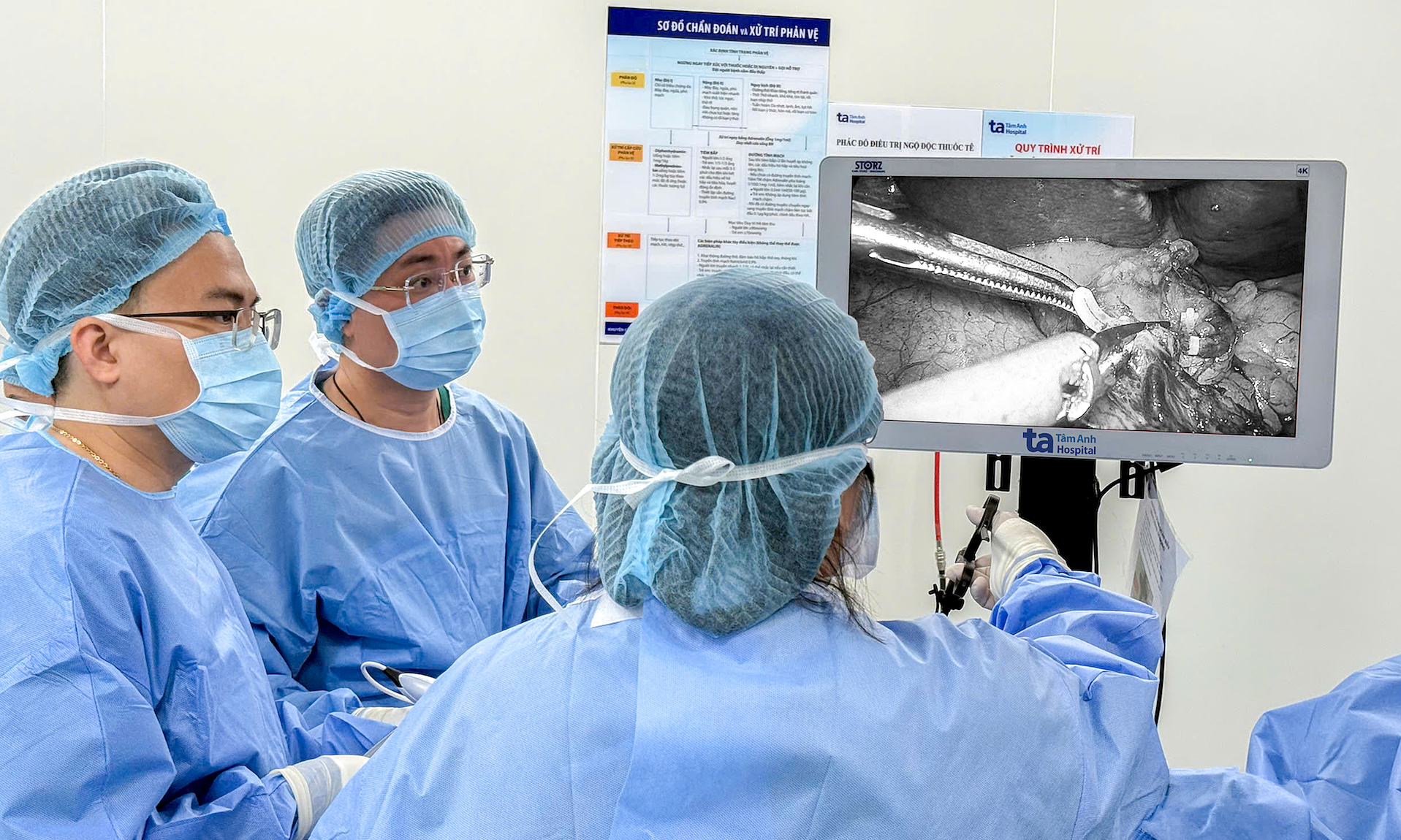
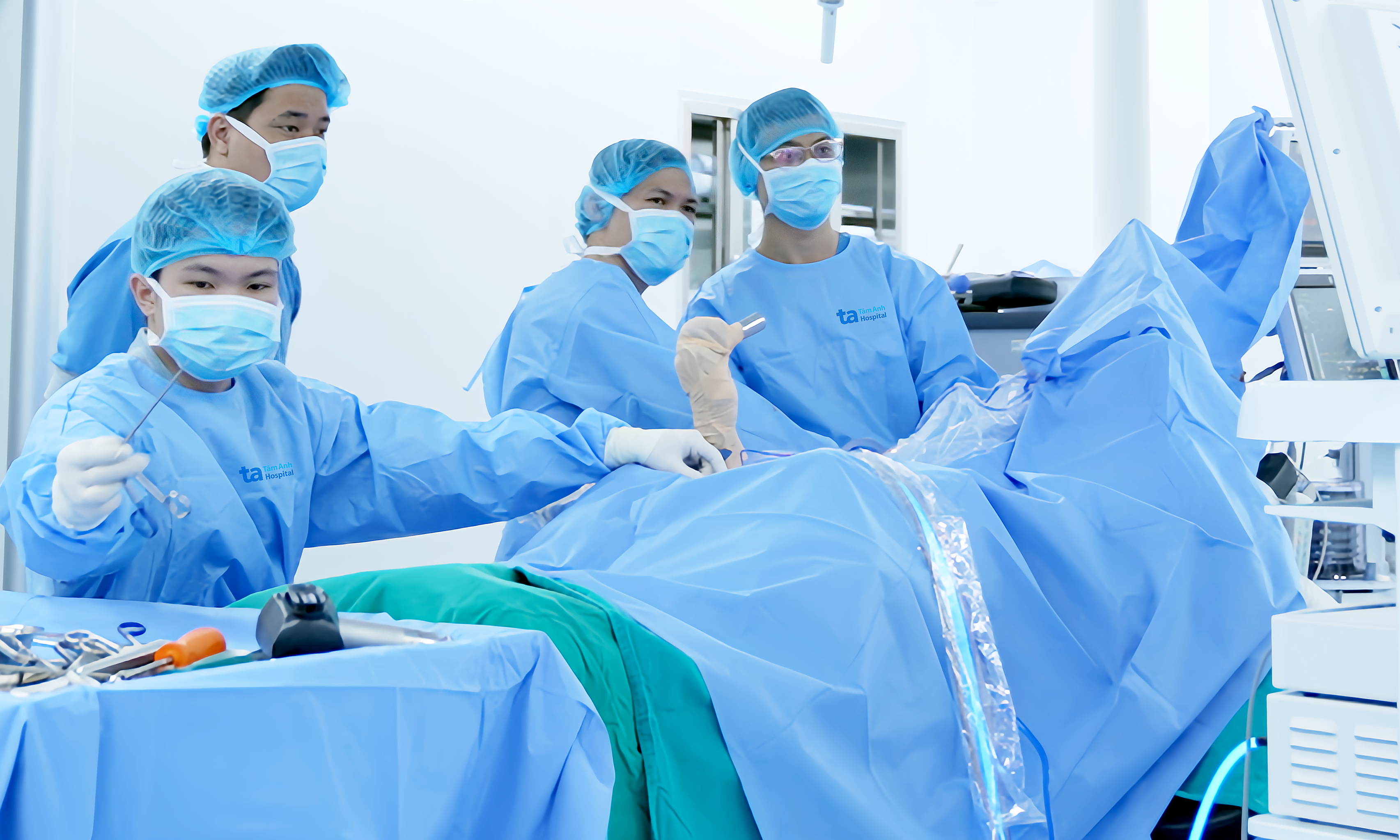
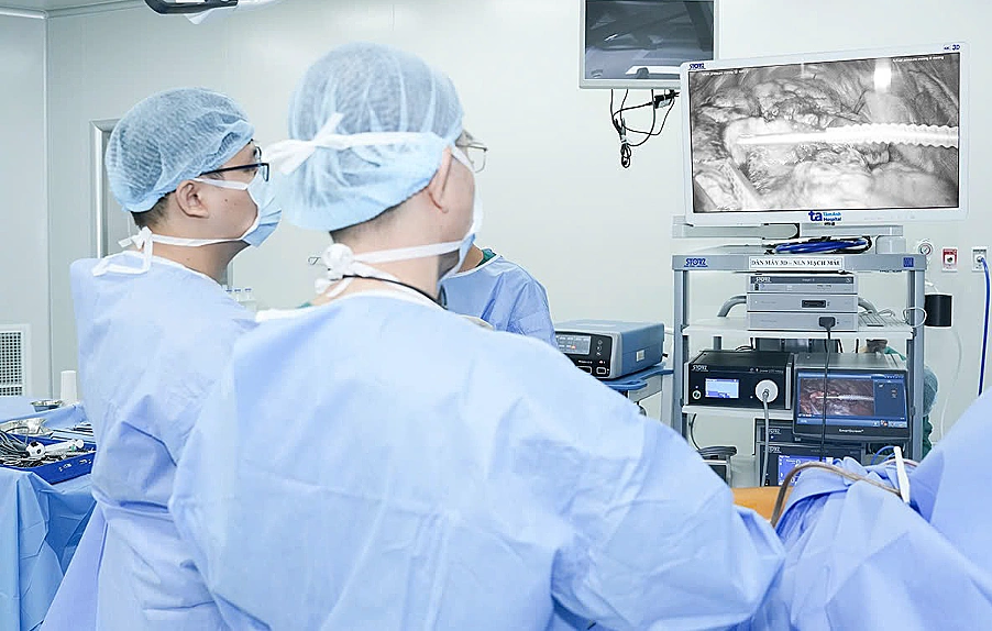
The patient was positioned at a 60-degree incline. The surgeon made three small skin incisions in the chest wall, inserting an endoscopic camera and specialized surgical instruments into the chest cavity. Magnified images from the camera allowed the surgeon to observe easily and precisely remove the tumor.
After 30 minutes, the surgeon removed a portion of the lung lobe containing the two lung nodules for a frozen section biopsy. While awaiting the results, the team proceeded to remove the thymus tumor. At this stage, the thymus tumor had not invaded adjacent organs, allowing the surgeon to dissect and remove the entire mass without affecting surrounding structures.
The frozen section biopsy confirmed both lung nodules were benign. Post-operative histopathology results identified the thymus tumor as type B2 thymic carcinoma.
 |
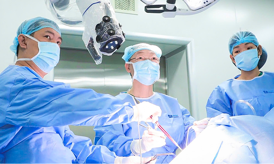
Doctors performing surgery to remove the thymus tumor and lung nodules. *Photo: Tam Anh General Hospital* |
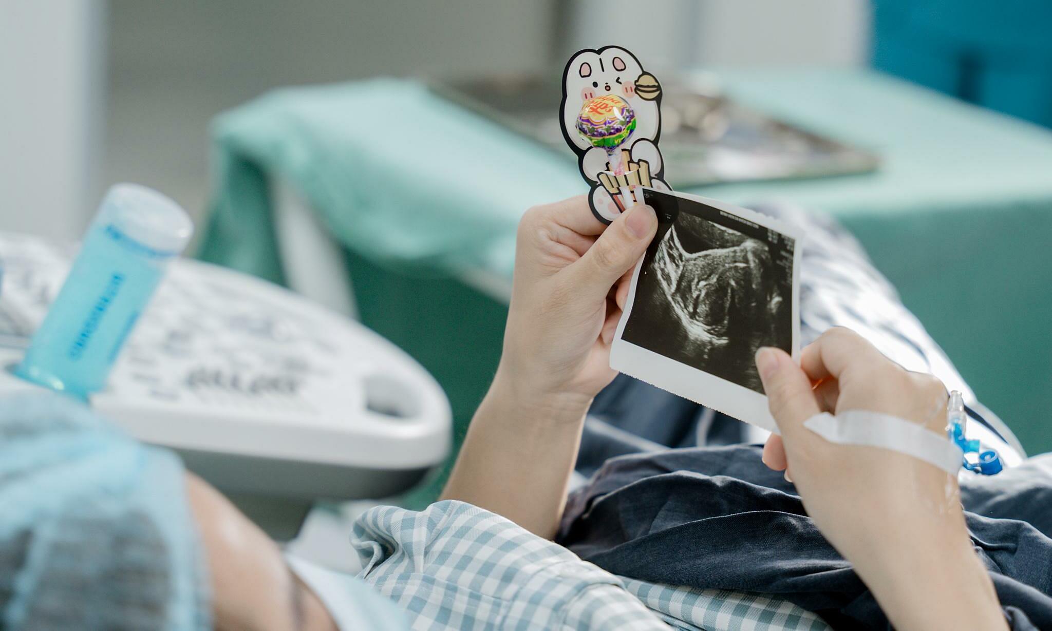
Post-operatively, Mrs. Chi's chest pain and shortness of breath resolved within a few hours, and she continued her treatment at the Oncology Center.
The thymus gland, located in the chest beneath the breastbone, is an organ of the lymphatic system within the immune system. Its role is to produce white blood cells, helping the body fight infections. Cancer develops when malignant cells appear on the surface of the thymus.
Doctor Phan Vu Hong Hai from the Center for Thoracic and Vascular Surgery at Tam Anh General Hospital Ho Chi Minh City noted that most patients with thymus tumors do not exhibit symptoms. Non-specific signs such as a persistent cough, shortness of breath, chest pain, difficulty swallowing, poor appetite, or weight loss are easily mistaken for other conditions, leading to delayed examination and late detection.
Doctor Hai recommends that adults undergo a regular health check-up at least once a year. Individuals aged 50-80 with a smoking history of 20 pack-years (one pack per day for 20 years or two packs per day for 10 years), or who are current smokers, should undergo screening for lung and thymus tumors as advised by a doctor. If symptoms such as chest pain, shortness of breath, or an unexplained cough persist for more than two weeks, patients should seek medical examination at a hospital.
Thu Ha
*Patient's name has been changed*
| Readers can submit questions about cancer here for doctors to answer |