A CT scan of the chest with vascular reconstruction at Tam Anh Hanoi General Hospital revealed that Hoang had a lung parenchyma region measuring approximately 91x99 mm, almost half the length of an adult lung (24 cm), occupying nearly the entire lower left lobe. This lung region was directly supplied by an arterial branch originating from the abdominal aorta.
Doctor Pham Thi Le Quyen, Deputy Head of the Respiratory Department at Tam Anh Hanoi General Hospital, diagnosed Hoang with pulmonary sequestration. This condition involves lung tissue developing separately, lacking normal respiratory function, and causing bronchiectasis and infection.
Doctor Quyen explained that pulmonary sequestration is a region of lung tissue that forms abnormally during the fetal period. Unlike normal lungs, this tissue region does not connect to the bronchial system. Because it is not ventilated and drains secretions normally, the sequestered lung region easily becomes a site for fluid stagnation, leading to recurrent infections, bronchiectasis, or lung abscesses.
The artery supplying the sequestered lung belongs to a high-pressure circulatory system. The continuous flow of high-pressure blood into these abnormal vessels, which have thin walls and incomplete structures, makes them prone to dilation, aneurysms, or rupture. Consequently, some patients may experience hemoptysis (coughing up blood), and less commonly, severe bleeding. This characteristic increases the risk of bleeding during surgery if the abnormal blood vessels are not carefully identified and controlled.
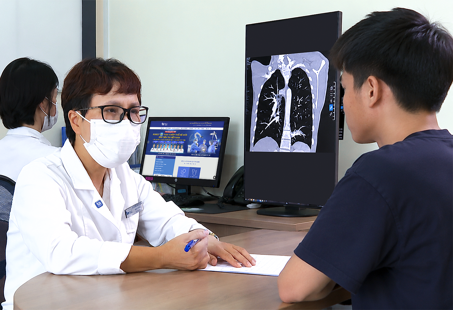 |
Doctor Quyen is consulting with the patient. Photo: Tam Anh General Hospital |
Doctor Quyen is consulting with the patient. Photo: Tam Anh General Hospital
Doctor Quyen noted that the case was complex because the sequestered lung region was large, occupying almost the entire lower left lobe and intermingled with healthy lung parenchyma, lacking clear boundaries. The prolonged infection had caused bronchiectasis, damaging the lung structure. If only a small portion were resected, diseased tissue might remain and continue to cause infection.
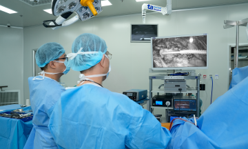
Following a multidisciplinary consultation, doctors decided to perform a lower left lobectomy using thoracoscopic surgery. With the aid of a high-resolution endoscopic system, surgeons carefully dissected and safely controlled the arterial branch supplying the sequestered lung before resecting the damaged lung lobe. This pre-operative vascular control helped limit the risk of bleeding while preserving as much healthy lung parenchyma as possible.
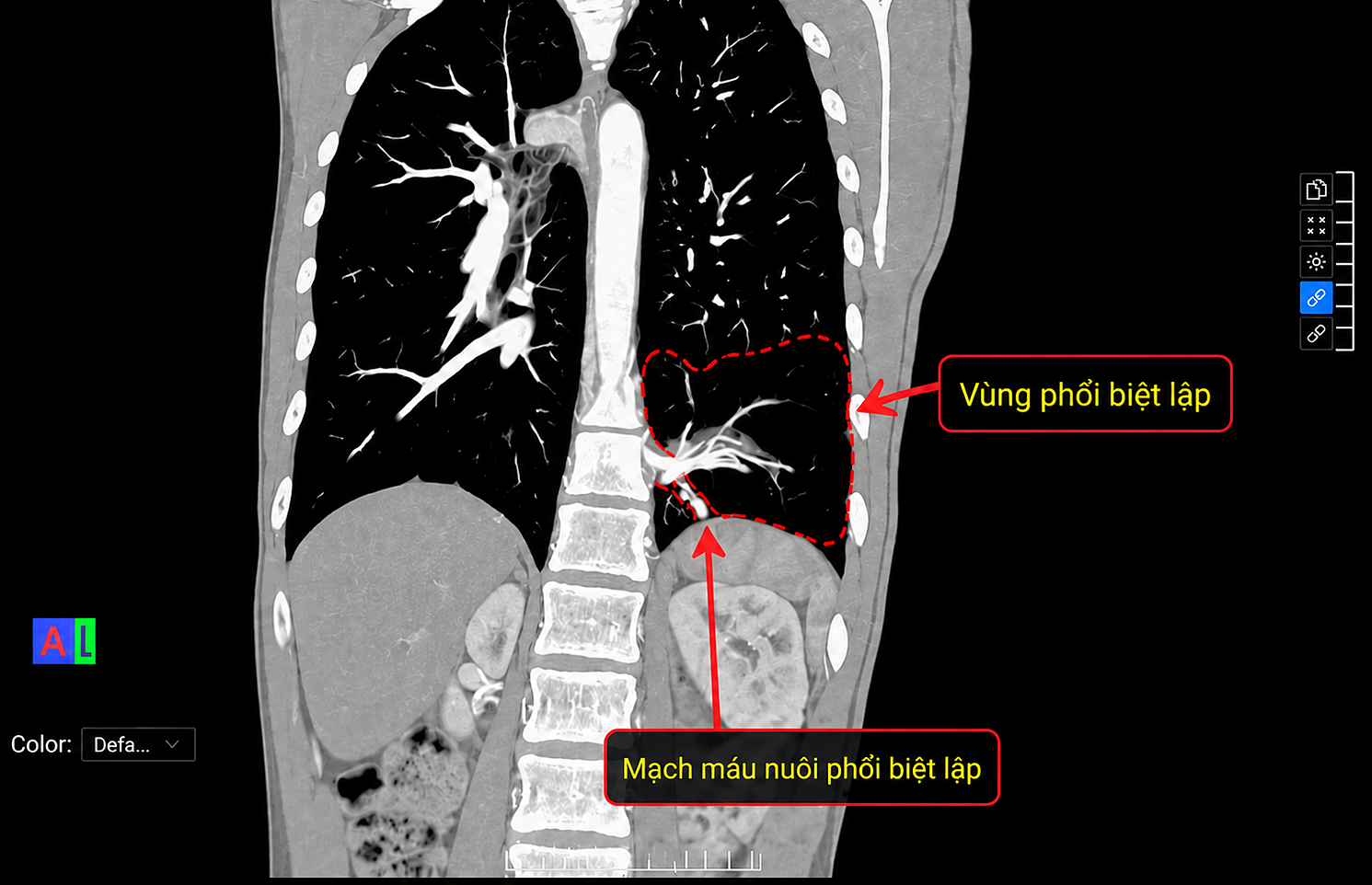 |
A CT scan of the lung showing the sequestered lung region in the lower left lobe, with the artery supplying it originating from the abdominal aorta. Photo: Tam Anh General Hospital |
A CT scan of the lung showing the sequestered lung region in the lower left lobe, with the artery supplying it originating from the abdominal aorta. Photo: Tam Anh General Hospital
After surgery, the patient's respiratory status, pleural drainage, pain control, and rehabilitation were monitored. After one week, the patient recovered well, experienced minimal pain, had the pleural drain removed, and was discharged. At a follow-up appointment more than two weeks later, the surgical incision had healed well, the remaining lung was well-ventilated, and there were no signs of acute infection.
According to Doctor Quyen, pulmonary sequestration accounts for approximately 0.15% to 6.4% of congenital lung diseases. The intralobar type, located within the same pleural sac as normal lung parenchyma, may not be detected at birth. Many cases are only diagnosed in adolescence or adulthood following recurrent respiratory infections, persistent cough and sputum production, or hemoptysis.
For symptomatic pulmonary sequestration or cases causing recurrent infections, surgery is typically the primary treatment method. In some instances, embolization may be considered.
Hoang Duong
| Readers can submit questions about respiratory diseases here for doctors to answer. |