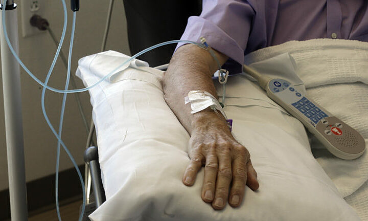
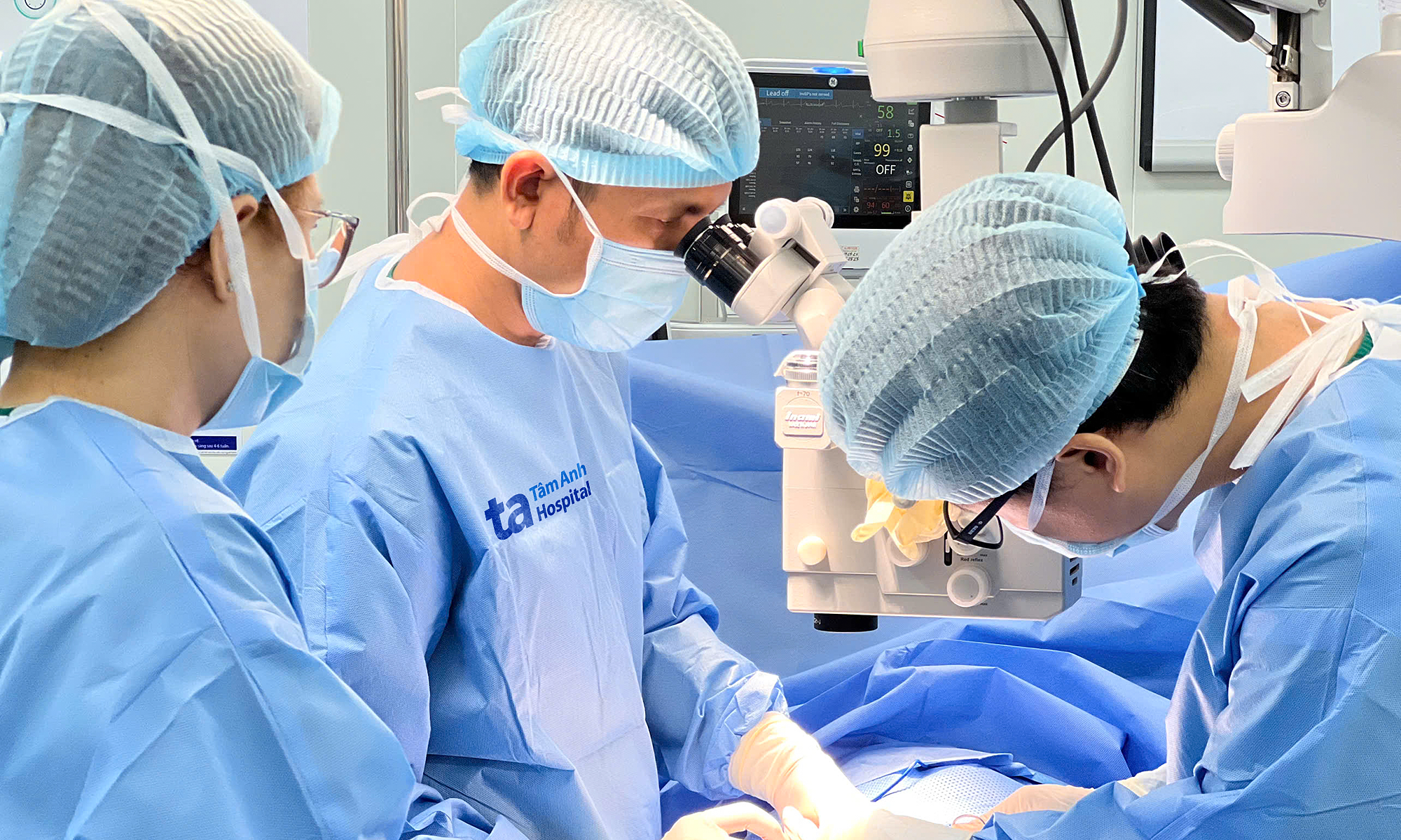
A computed tomography (CT) scan at Tam Anh General Hospital Ho Chi Minh City revealed a right kidney angiomyolipoma in Loan, measuring approximately 7.1x7.2 cm and nearly 8 cm long, almost the size of a normal kidney. The tumor had ruptured, leading to hemorrhage and a perirenal hematoma.
Doctor of First Degree Specialist Duong Pham Van Thanh from the Emergency Department stated that the patient faced a significant risk of blood loss. Delayed or inappropriate treatment could necessitate partial or complete removal of the kidney to control the bleeding.
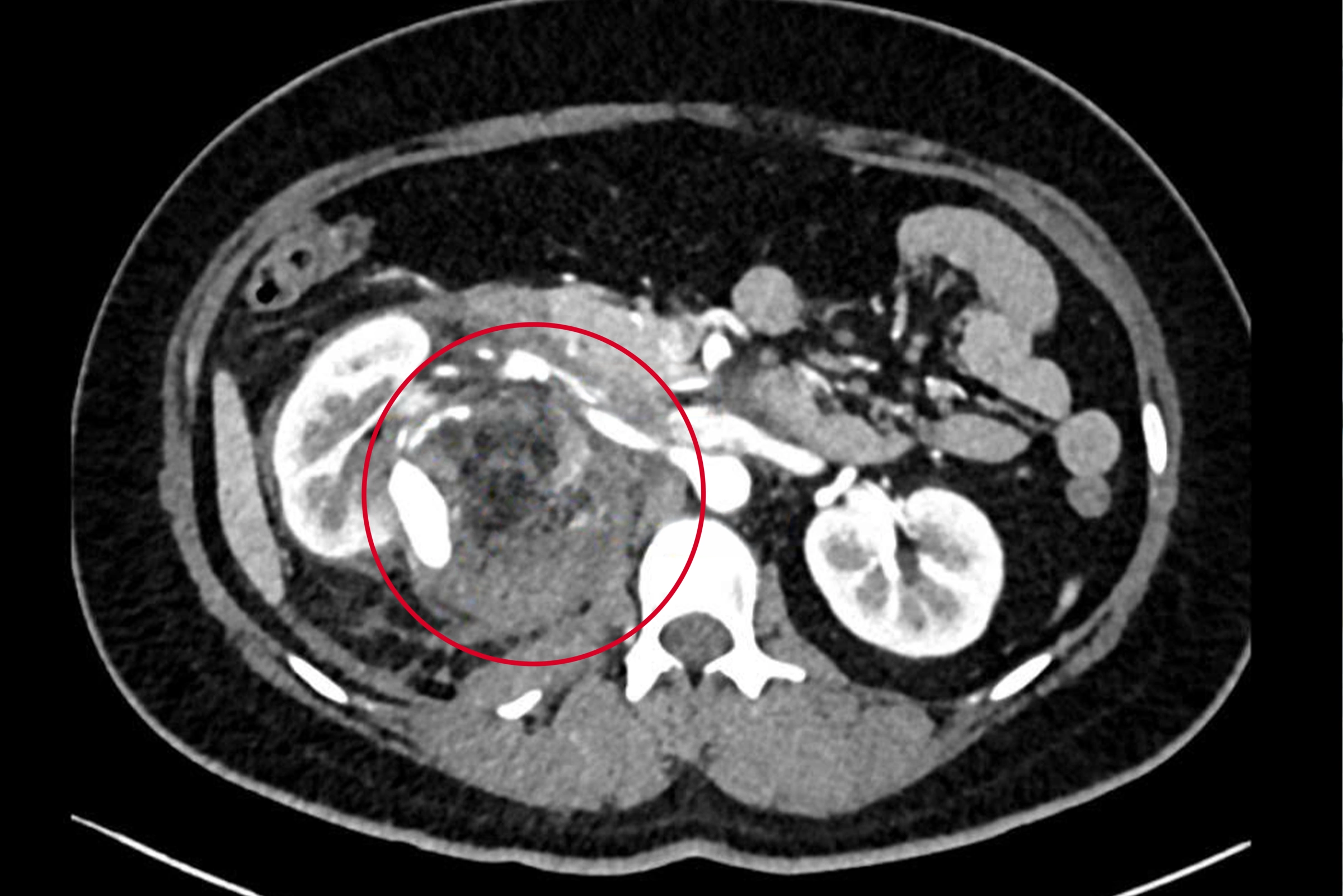 |
The tumor (red circled area) is almost the size of a kidney. Photo: Tam Anh General Hospital |
A multidisciplinary team, comprising specialists from the Emergency Department, Urology - Nephrology - Andrology Center, and Imaging Diagnosis and Interventional Radiology Center, convened. They opted against immediate surgery, choosing instead an emergency endovascular embolization to control bleeding, with the primary goal of preserving the patient's kidney.
Doctor of First Degree Specialist Nguyen Trung Duc from the Imaging Diagnosis and Interventional Radiology Center explained that the tumor was fed by numerous arterial branches, one of which contained a large pseudoaneurysm, identified as the source of the hemorrhage. Operating at this stage would have carried a high risk of severe bleeding.
The team performed the procedure using a digital subtraction angiography (DSA) system. Through a small puncture in the femoral artery, doctors guided a microcatheter to the arterial branches feeding the tumor. Embolization material was then used to completely seal the pseudoaneurysm and the bleeding vessels.
Following the intervention, the hemorrhage was successfully controlled, and Loan's right kidney was preserved. She recovered well and was discharged the next day.
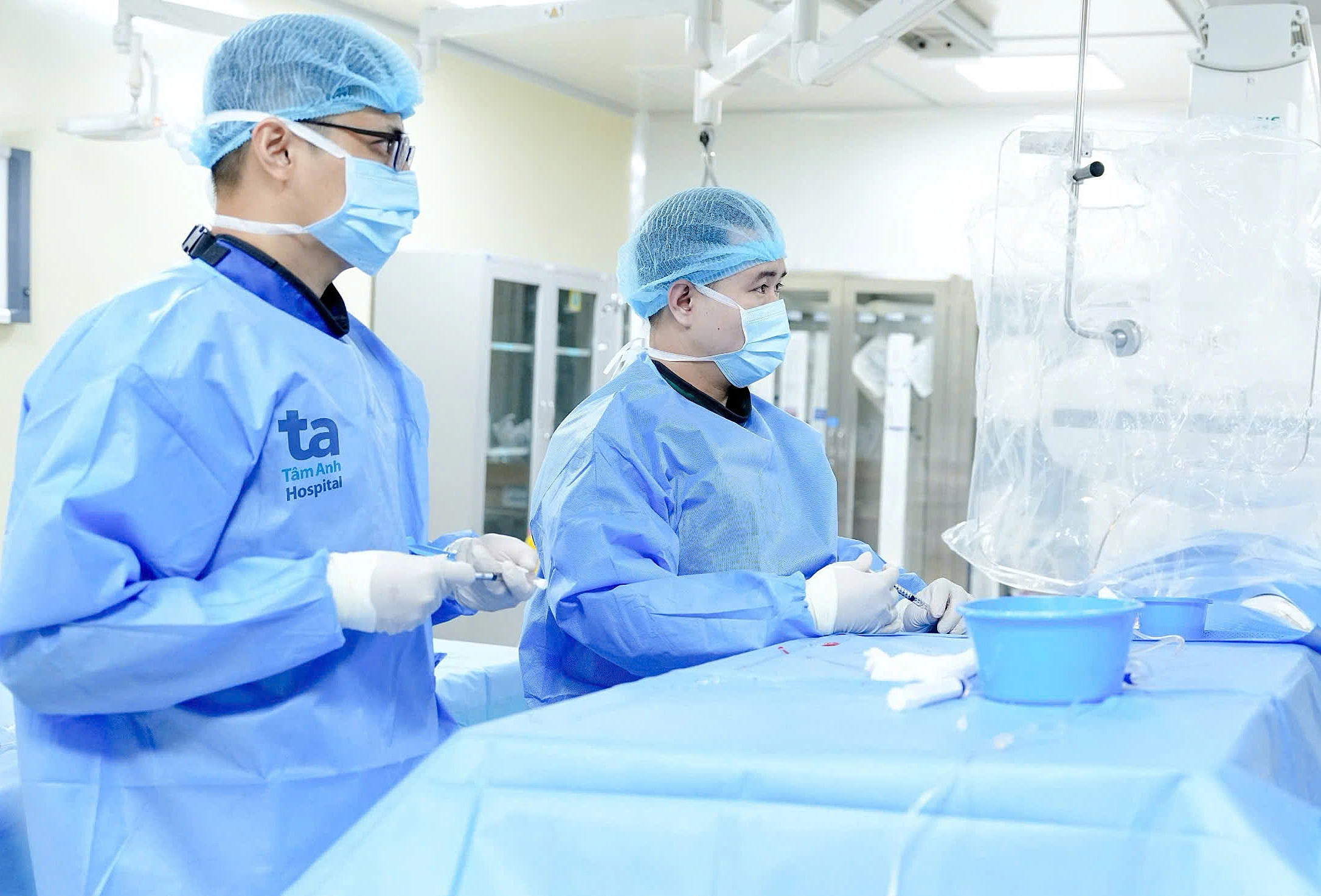 |
Doctor Duc (right) and the team performed the intervention to seal the pseudoaneurysm and the vessels supplying the tumor for the patient. Photo: Tam Anh General Hospital |
According to Doctor Duc, renal angiomyolipoma is a common benign kidney tumor, resulting from the abnormal growth of vascular, smooth muscle, and adipose tissues. Most cases are asymptomatic, often discovered incidentally during routine health check-ups or after complications arise.
When renal angiomyolipoma grows large, becomes symptomatic, or develops vascular abnormalities like aneurysms or pseudoaneurysms within the tumor, the risk of rupture and hemorrhage significantly increases. This can lead to perirenal or retroperitoneal bleeding, causing severe blood loss and potentially becoming life-threatening without prompt intervention.
Historically, ruptured renal angiomyolipoma causing hemorrhage often mandated emergency surgery, frequently involving partial or total nephrectomy to control bleeding. Today, selective endovascular intervention offers a less invasive option. This technique helps stop bleeding, minimizes damage to healthy kidney tissue, and enhances the likelihood of preserving kidney function. Post-treatment, patients require regular follow-up and imaging to monitor for recurrence or the need for further intervention.
Patients experiencing symptoms such as persistent flank pain, worsening abdominal discomfort, hematuria, or unexplained fever should seek medical attention promptly. Doctors can utilize various imaging diagnostic techniques to detect the condition and intervene before complications arise.
Nhu Ngoc
* The patient's name has been changed