Answer:
Bilateral fetal ventriculomegaly is a condition where the cerebral ventricles dilate. This can result from cerebrospinal fluid obstruction within the brain's cavities, disrupting fluid flow, or from structural brain abnormalities. The condition is categorized into mild (10-12 mm), moderate (13-15 mm), and severe (over 15 mm) levels.
Ventriculomegaly can manifest as an isolated defect or be linked to central nervous system abnormalities, such as agenesis of the corpus callosum or neural tube defects. It may also be associated with genetic disorders, including Dandy-Walker, Down, Edwards, Patau, and Chiari syndromes. Furthermore, congenital infections like CMV, Toxoplasmosis, or Zika, cerebral hemorrhage, or a reduction in surrounding intracranial tissue volume are also risk factors for ventriculomegaly.
In your case, the 29-week ultrasound showed bilateral ventriculomegaly, but the severity remains unclear. A specialized fetal neuro-ultrasound is essential for doctors to thoroughly assess the development of the brain structure.
Additionally, doctors may order a fetal magnetic resonance imaging (MRI) scan to detect any lesions that ultrasound might miss. MRI utilizes magnetic fields and is safe for both the mother and the fetus. This method significantly aids in evaluating the structure of brain parenchyma layers, sulcal formation, lesions resulting from abnormal neuronal migration, and damage related to hemorrhage.
 |
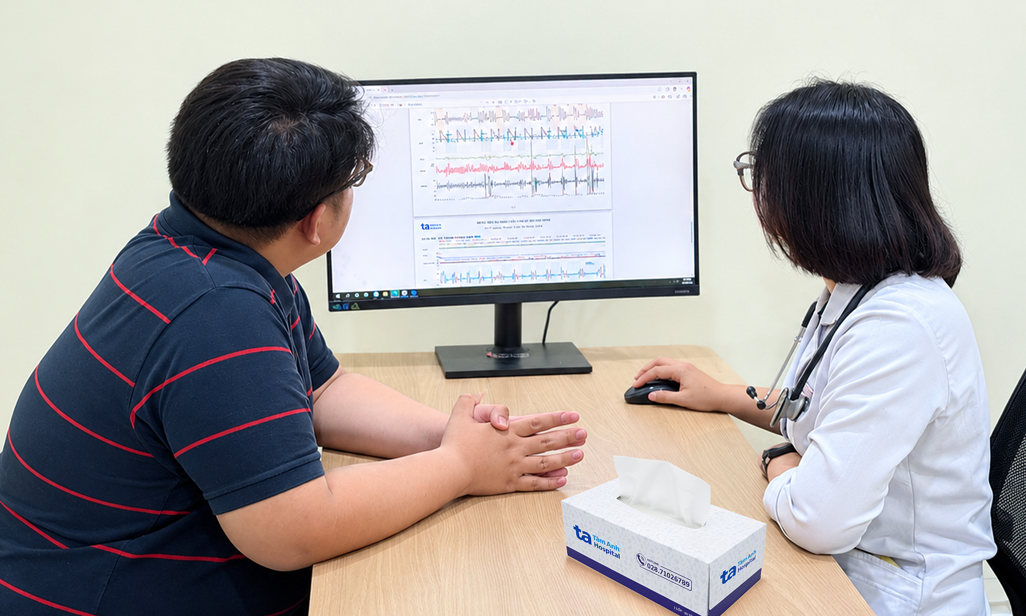
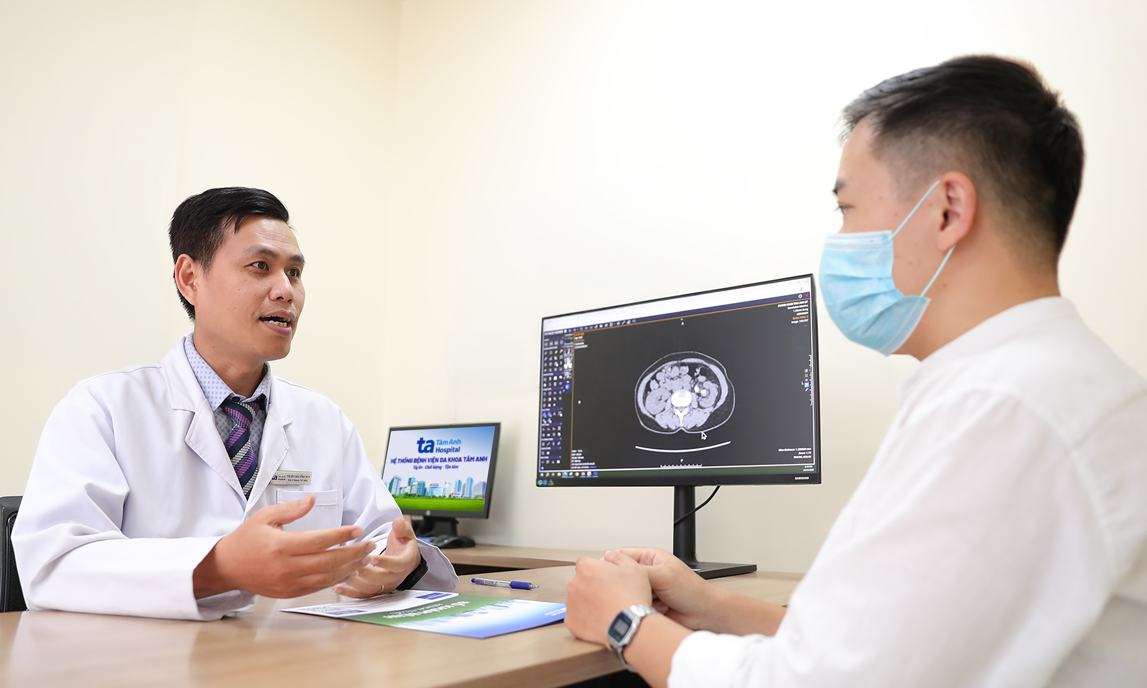
Dr. Nguyen Hoang Long consults a pregnant woman. Illustration: Tam Anh District 7 General Clinic. |
When fetal ventriculomegaly is present, amniocentesis is an essential procedure to determine if the cause is related to genetic disorders or an infection. Guided by an ultrasound system, doctors insert a thin needle through the pregnant woman's abdominal wall and uterine wall into the amniotic sac, extracting a sufficient amount of amniotic fluid for genetic testing.
Furthermore, you should undergo regular scheduled ultrasounds to monitor the progression of ventricular size and the fetus's overall development. Examinations at specialized fetal medicine units, with close interdisciplinary coordination, advanced neuro-ultrasound systems, and modern fetal MRI systems, provide a comprehensive assessment of fetal development. This enables doctors to advise on appropriate monitoring and intervention plans for each stage of pregnancy and prepare the safest delivery options for both mother and baby.
Dr. Nguyen Hoang Long
Fetal Medicine Unit
Tam Anh District 7 General Clinic
| Readers can submit questions about obstetrics and gynecology here for doctors to answer. |