Doctor Dao Duy An Duy, from the Orthopedic Trauma Center at Tam Anh General Hospital, TP HCM, reported that Ms. Nhan's complex fracture was the first of its kind seen at the hospital. He explained that the femoral neck connects the femoral head to the shaft, while the intertrochanteric region is a transitional area. Despite their proximity, these hai sites have distinct structures and load-bearing characteristics, typically resulting in separate fractures with different treatment approaches.
Ms. Nhan's fractures at both sites in the upper femur led to deformation of the hip joint and femur, bone displacement, and loss of anatomical landmarks. After consultation, doctors opted for a "hai-in-one" major surgery. This involved an artificial hip replacement and greater trochanter bone fixation with cable plating, all performed in a single operation.
Doctor Duy noted the surgery's increased complexity because the upper femur had fractured into multiple pieces, complicating the artificial joint placement. Despite this, it was considered the optimal approach to reduce pain, facilitate rapid recovery, and mitigate complications from extended immobility.
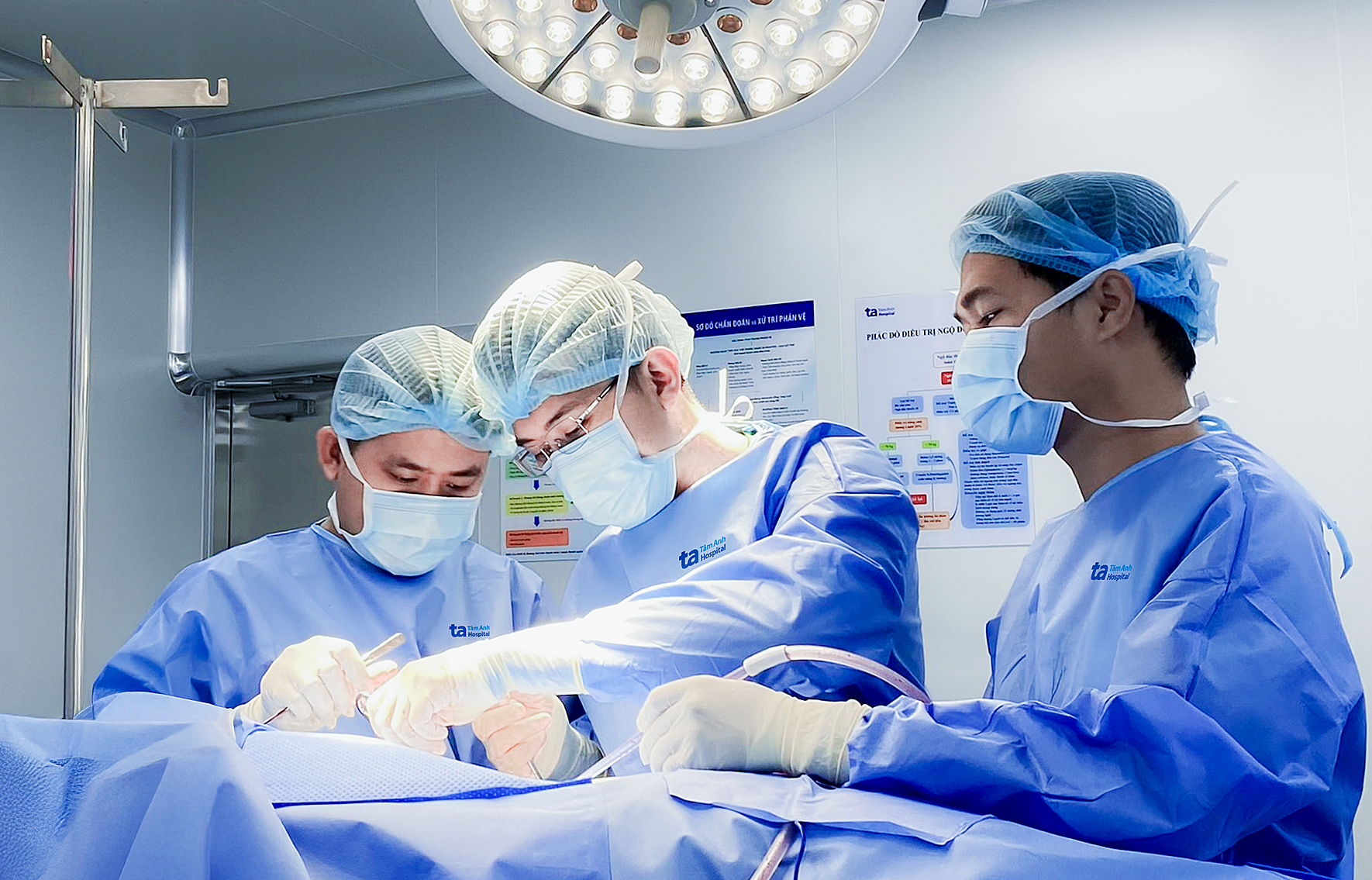 |
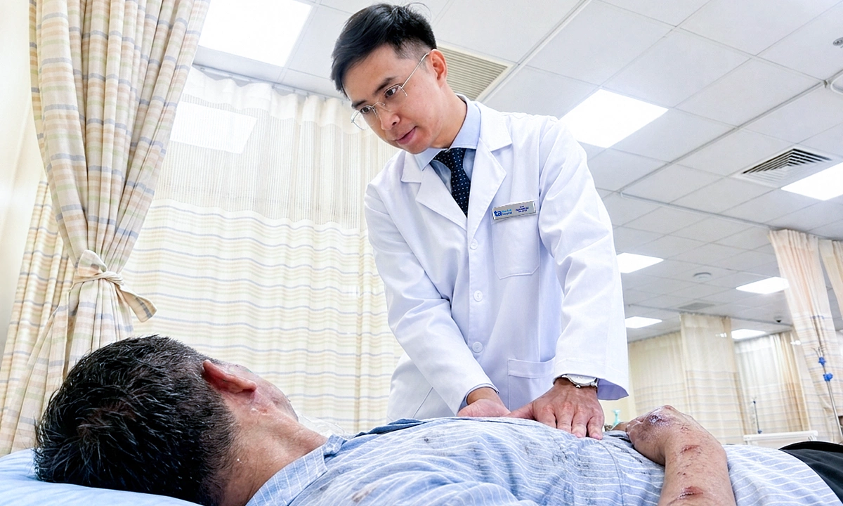
Doctor Duy (center) performs joint replacement and bone fixation for Ms. Nhan. *Photo: Tam Anh General Hospital* |
Doctors utilized TraumaCAD software to select the optimal joint type, placement, and cable plate length for the patient. Ms. Nhan received a total artificial hip replacement through a posterior approach, which allowed for clear visualization and reduction of the fracture. Following this, the fractured bone was addressed by applying a plate near the trochanteric region and femoral shaft. Metal wires were then tightened around the bone to secure large fragments, restoring anatomical structure and muscle attachment points.
For complex fractures like Ms. Nhan's, post-operative rehabilitation is crucial. Exercise regimens ensure adequate time for bone healing while promoting early patient mobilization to prevent complications from prolonged bed rest, including pressure ulcers, pulmonary embolism, and urinary tract infections.
For the first 6 weeks, Ms. Nhan used a walker or crutches, with limited weight-bearing on her left leg. The following 6 weeks involved walking without assistive devices. Doctor Duy forecasts a full recovery and a return to normal life within ba months.
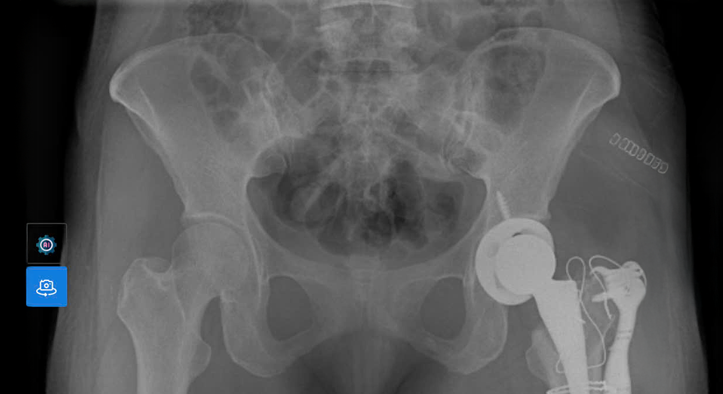 |
X-ray of Ms. Nhan's hip joint after treatment. *Photo: Tam Anh General Hospital* |
Doctor Duy highlighted that modern imaging equipment, including a 1975-slice CT scanner and ba Tesla MRI, alongside planning software, supported doctors in making precise decisions for complex orthopedic trauma. This advanced technology allowed patients to be quickly relieved of pain and regain mobility within mot-hai days post-surgery.
Phi Hong
*Patient's name has been changed
| Readers can send questions about musculoskeletal diseases here for doctors to answer. |