The device was researched and developed by Doctor Dinh Thi Hoang Anh, Head of the Ophthalmology Department, Hong Ngoc General Hospital, in collaboration with Professor Kalinnikov Yury, an ophthalmologist from the Russian Federation. The invention has been exclusively registered in Russia.
According to Doctor Hoang Anh, this is a practical support tool for pupil reconstruction surgery. This tool is used when the eye sustains severe damage, leading to deformed or widely dilated pupils, which significantly affects visual quality.
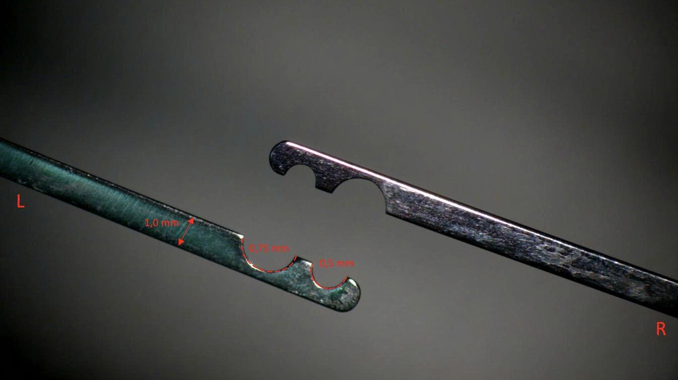 |
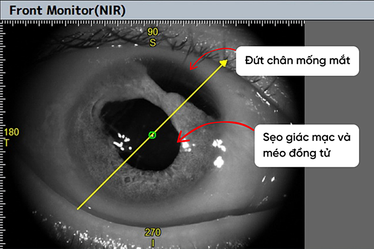
The device helps measure precise pupil size directly on the pupil, unlike previous methods that measured on the cornea. Photo: Hospital provided |
Treating physicians can directly measure the size at the pupil plane inside the eye, rather than estimating it on the corneal surface as was previously done. This provides a better basis for controlling the roundness, symmetry, and precise size of the pupil during reconstruction.
The hospital has applied the new device in several recent surgeries. For example, patient Ha Thanh Thuy, 54, from Hung Yen, suffered a work accident in late 2025 when metal fragments hit his eye. Despite undergoing multiple previous interventions, including corneal suturing, cataract removal, and vitrectomy, his vision had barely improved. Upon arriving at Hong Ngoc Eye Center, the patient still experienced blurred vision, double vision, light sensitivity, and significantly impacted daily life.
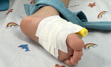
Upon examination, doctors identified severe multiple traumas in the patient's right eye: deep central corneal scarring, iris root detachment, and a distorted, dilated pupil. Many important structures in the patient's eye were damaged simultaneously.
 |
The patient's right eye showed severe damage after the accident. Photo: Hospital provided |
"The goal of treatment is not only to restore anatomy but also to improve visual function", Doctor Hoang Anh said.
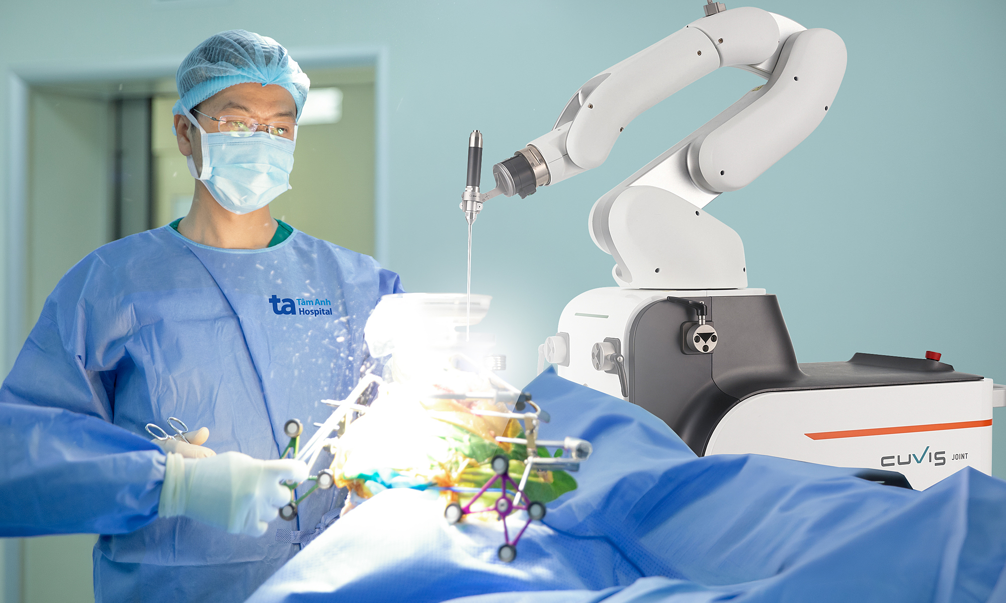
The surgical team decided to perform a combined multi-technique surgery in a single session: iris-fixated intraocular lens implantation, pupil reconstruction, and penetrating keratoplasty. A significant challenge was that the patient's eye had lost important supporting structures like the lens and vitreous. When dissecting the cornea for grafting, the eyeball was in an "open" state, easily leading to severe complications such as expulsive hemorrhage, directly threatening vision. Expulsive hemorrhage is a dangerous ophthalmic complication, causing pressure that can rupture the eyeball, expelling all intraocular contents and severely impacting the patient's vision.
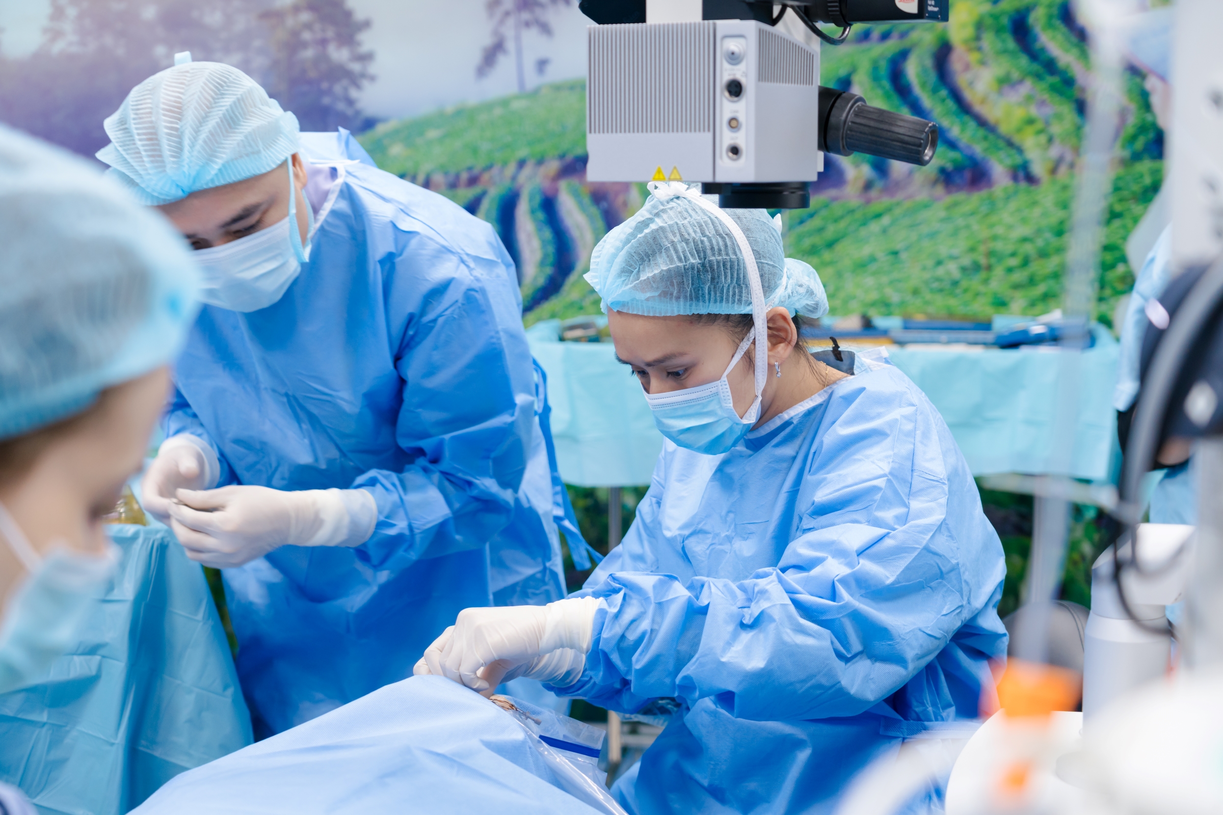 |
The difficult surgery involved doctors using the new device for treatment. Photo: Hospital provided |
To reduce risks, the doctors carefully planned the surgical sequence. First, they performed iris-fixated intraocular lens implantation and iris reconstruction to re-establish intraocular structures. Then, the pupil was reduced to about 1,5 mm to minimize optical aberrations and improve vision. The pupil measurement device aided in directly determining the size at the pupil plane, allowing for more precise manipulation. Once the intraocular structures were stable, the team proceeded with penetrating keratoplasty.
After surgery, the cornea was clear, the surgical incision was sealed, the pupil was reconstructed to the correct size, and the intraocular lens was stable. The patient's vision improved from counting fingers at 0,5 m to 5/10. The outcome not only helped the patient improve vision but also demonstrated the potential application of the pupil measurement device in complex ophthalmic cases.
Van Ha