Mr. Hung had a long history of chronic obstructive pulmonary disease, type 2 diabetes, hypertension, and dyslipidemia. Approximately six months prior, he experienced increased shortness of breath, attributing it to a worsening of his chronic obstructive pulmonary disease and continuing his medication. A computed tomography (CT) scan at a local medical facility revealed several small, scattered nodules in both lungs. He then visited Tam Anh General Hospital Hanoi for a positron emission tomography (PET/CT) scan, which showed similar results. Notably, a solid nodule in the lower lobe of his right lung, measuring approximately 13x15x10 mm, exhibited increased fluorodeoxyglucose (FDG) uptake, a potential indicator of a malignant lesion.
According to Master of Science, Doctor, Level II Specialist Nguyen Le Vinh, Head of Thoracic Surgery at the Center for Cardiovascular and Thoracic Surgery, the lesion was suspected to be an early-stage lung tumor. Surgery was recommended to accurately determine the tumor's nature and ensure its complete removal.
The medical team opted for a thoracoscopic lung nodule biopsy. This minimally invasive technique involves creating small incisions, which are less invasive than open surgery, thereby reducing pain and blood loss. Consequently, patients with chronic obstructive pulmonary disease face a lower risk of postoperative atelectasis, pneumonia, and respiratory failure.
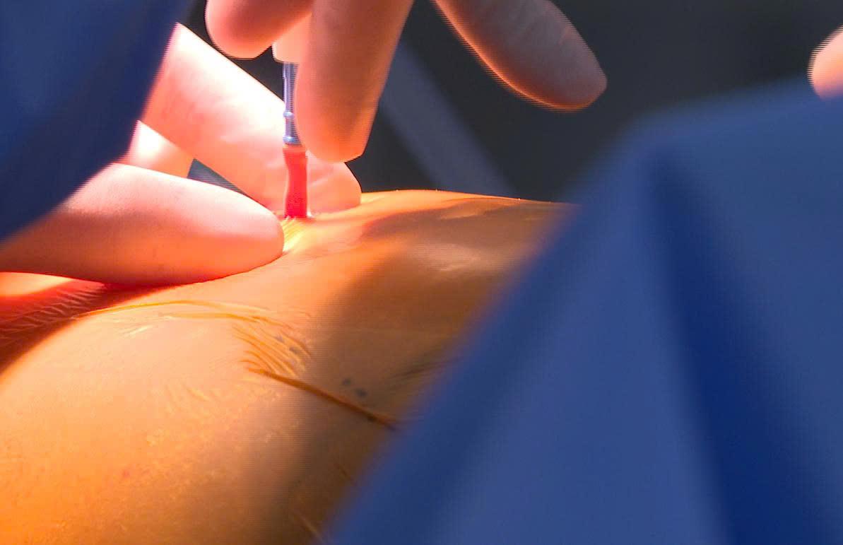 |
Surgeons perform thoracoscopic surgery on Mr. Hung through small, minimally invasive incisions. *Photo: Tam Anh General Hospital*
The surgeons performed a wedge resection, removing a small portion of the lung containing the lesion, to preserve healthy lung tissue in case the lesion was not malignant. After 15 minutes, a frozen section biopsy confirmed the tumor was malignant. The team then proceeded with a complete right lung lobectomy and lymph node dissection.
The surgery was successful. Pathology results, combined with immunohistochemistry, confirmed the patient had keratinizing squamous cell carcinoma in the lung. The tumor measured approximately 1,6 cm and remained confined within the lung parenchyma, without invading other organs.
Following surgery, Mr. Hung received close monitoring, respiratory rehabilitation, blood sugar and blood pressure control, and nutritional management. He subsequently recovered well, experienced reduced shortness of breath, and was discharged.
According to Doctor Vinh, chronic obstructive pulmonary disease is a risk factor for lung cancer. Both conditions share several common risk factors, most notably smoking, air pollution, and exposure to dust and occupational chemicals. The prolonged chronic inflammation within the respiratory tract of individuals with chronic obstructive pulmonary disease can damage cellular deoxyribonucleic acid (DNA), promote gene mutations, and create a conducive environment for cancer development.
For early-stage lung cancer, lung lobectomy combined with lymph node dissection remains the standard treatment, increasing survival rates if the tumor is detected before it spreads. Doctor Vinh advises individuals with chronic obstructive pulmonary disease, particularly those over 50 years old with a history of smoking or prolonged past smoking, to undergo regular respiratory specialist examinations and lung cancer screenings.
If symptoms such as a change in cough characteristics, rapidly worsening shortness of breath, coughing up blood, persistent chest pain, or weight loss appear, patients should seek medical attention promptly.
Thanh Ba
| Readers can submit questions about respiratory diseases here for a doctor's response. |